Required section · Section 3 of 6
What each pattern supports, and what the screen cannot see
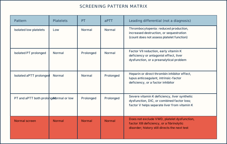
An isolated prolonged PT with normal aPTT supports a differential that includes reduced factor VII activity, an early vitamin K deficiency or vitamin K antagonist effect (factor VII has the shortest half-life of the vitamin K-dependent factors, so its reduction shows up first), liver dysfunction, or a preanalytical problem. An isolated prolonged aPTT with normal PT supports heparin or direct thrombin inhibitor effect, lupus anticoagulant, an intrinsic-pathway factor deficiency, or a factor inhibitor. A normal aPTT does not exclude mild hemophilia A or B; the test's sensitivity depends on the reagent and how mild the deficiency is.
When both PT and aPTT are prolonged, consider more severe vitamin K deficiency or antagonism, liver synthetic dysfunction (the liver makes most coagulation factors), or a consumptive process such as disseminated intravascular coagulation (DIC) in the right clinical setting. Factor V activity is one part of separating hepatic synthetic dysfunction from isolated vitamin K deficiency because factor V is not vitamin K dependent: a low result can support impaired synthesis rather than vitamin K effect alone, but can also occur with consumption or other causes and is not definitive by itself. DIC is a clinical-laboratory diagnosis: a falling platelet count, PT prolongation, rising fibrin-related markers, and a low or falling fibrinogen support it, but a normal or even increased fibrinogen does not exclude DIC, because fibrinogen behaves as an acute-phase reactant and can start high before it falls.
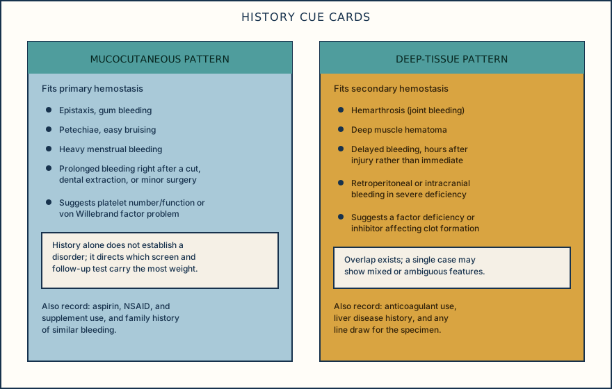
Hemophilia A or B commonly presents as a normal PT with a prolonged aPTT and a normal platelet count, but the screen alone cannot distinguish hemophilia A from a phenocopy; specific factor VIII or IX activity is required. A reduced factor VIII activity should also prompt evaluation for VWD, including type 2N VWD, before hemophilia A is assumed, because VWF normally protects factor VIII from clearance. For suspected VWD, the initial specific work-up is VWF antigen, platelet-dependent VWF activity, and factor VIII coagulant activity; a VWF level below 0.30 IU/mL supports type 1 VWD regardless of bleeding, and below 0.50 IU/mL supports it in a patient with abnormal bleeding. A VWF activity-to-antigen ratio below 0.70 is used to flag a type 2 VWD pattern for further multimer or collagen-binding classification. VWF is also an acute-phase reactant and rises with stress or inflammation, so testing at baseline health is preferred and an unexpected result may need repeating.
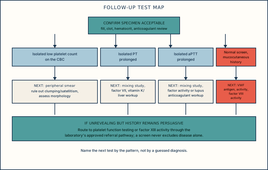
A closure-time test such as PFA can be abnormal in VWD or a severe platelet disorder, but it has limited sensitivity for mild VWD and mild secretion defects, so a normal result cannot exclude either. Only after specimen quality and anticoagulant exposure are resolved should a 1:1 mixing study be interpreted: in a drug-free specimen, correction toward the laboratory's criterion supports a factor deficiency, while failure to correct can reflect an inhibitor. A DOAC-affected mix cannot be read by that pattern because the drug can alter the clot-based result; use the local medication-interference approach before assigning an inhibitor or deficiency. Immediate correction followed by prolongation after incubation supports a time-dependent inhibitor pattern only after drug interference is excluded. When factor activity is needed, state whether the local method is one-stage clot-based or chromogenic and interpret it by that method; the two are not interchangeable. Coagulation specimens require the validated sodium-citrate tube and blood-to-citrate ratio; underfill or an uncorrected high hematocrit falsely prolongs clot-based results, and heparin contamination from a line draw can mimic an inhibitor pattern on aPTT.
A rare but real limitation: factor XIII deficiency can cause bleeding with a fully normal PT and aPTT, because factor XIII acts after fibrin has already formed and neither screening test detects its loss. Factor XIII activity is the targeted study when the pattern and history point there, and a rare-factor diagnostic algorithm goes beyond this pattern discussion; route that decision through your laboratory's referral pathway.
Exact reference intervals, reagent and analyzer combinations, mixing-study cutoffs, D-dimer category mapping for a DIC score, and critical values are set locally and verified per your laboratory's procedure; nothing above should be read as a portable interval. Name the pattern, name what it does and does not exclude, then pick the one test that actually narrows it further.
Illustrative drawing — this picture was drawn rather than captured.
Illustrative drawing — this picture was drawn rather than captured.
Illustrative drawing — this picture was drawn rather than captured.
Knowledge checks
Reading and checks are open. Sign in only to save.
Knowledge check 1
Knowledge check 2
Knowledge check 3
Knowledge check 4
Section status
Finish this section
Reading and checks are open. Sign in only to save.
The module finishes after every required section is marked done and every check in those sections is correct.