Required section · Section 6 of 6
Debrief: supported pattern and boundaries
This case supports persistent true multilineage elevation and a PV-focused integrated workup. It does not establish PV, exclude all reactive contributors, or demonstrate PMF morphology. The diagnosis and treatment decisions remain outside the laboratory result release.
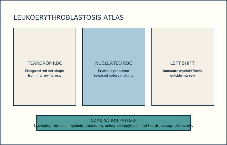
ICC separates prefibrotic PMF with fibrosis below grade 2 from overt PMF with reticulin or collagen fibrosis grade 2 or 3. Overt PMF can show nucleated red cells, myeloid left shift, anisopoikilocytosis, and teardrop erythrocytes on the film. Fibrosis grade is a bounded marrow-pathology task: a qualified reviewer assesses an adequate marrow reticulin/collagen stain using the controlled grading procedure; it is not assigned from the blood film.
Molecular reports should identify specimen, method, target or variant, analytical sensitivity, result, and method limitations. The laboratory validates its own limit of detection, covered variants, and reportable range. Release only what the validated method measures and name the local policy or morphology dependency.
Illustrative drawing — this picture was drawn rather than captured.
Knowledge checks
Reading and checks are open. Sign in only to save.
Knowledge check 1
Knowledge check 2
Section status
Finish this section
Reading and checks are open. Sign in only to save.
The module finishes after every required section is marked done and every check in those sections is correct.