Required section · Section 3 of 6
Reading the Cast Against the Rest of the Result Set
A hyaline cast alone tells you almost nothing. It is low-refractile uromodulin matrix, it can increase with dehydration, fever, or strenuous exercise, and it must be read together with urine concentration, chemistry, cells, and renal function before it earns any interpretive weight. Do not let a hyaline cast anchor a laboratory comment by itself.
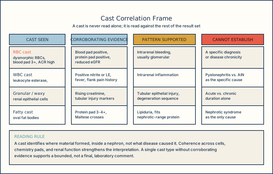
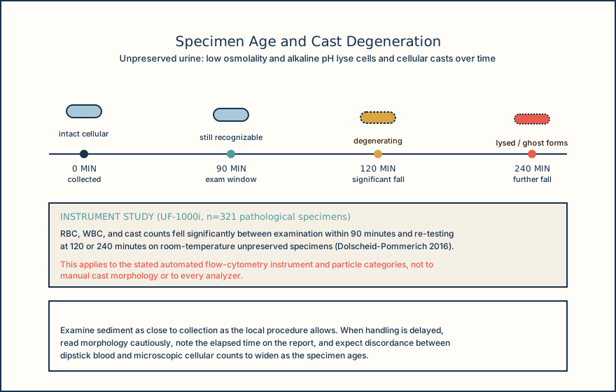
An RBC cast is different in weight but not in the need for correlation. It supports bleeding within the nephron, usually a glomerular pattern, and it is one of the more specific casts you will see. It is still not a final diagnosis: RBC casts occur in biopsy-proven acute interstitial nephritis as well, so the glomerular association has real exceptions. Dysmorphic RBCs and acanthocytes add support for glomerular hematuria, but published detection methods and cutoffs vary enough that no universal laboratory interpretation rule exists; phase-contrast microscopy improves visualization of dysmorphic RBCs and hyaline casts, and bright-field findings cannot be treated as interchangeable with phase-contrast findings without local validation. A related specimen-quality point: the cell-free blood pad on a reagent strip measures heme peroxidase activity, and that activity can persist after RBCs have already lysed. A strongly positive blood pad with few intact RBCs on microscopy is a specimen-quality flag, not a contradiction to ignore.
A WBC cast supports an intrarenal inflammatory pattern and occurs with pyelonephritis, acute interstitial nephritis, and some nephritic processes. It is insensitive for acute interstitial nephritis, meaning its absence does not argue against that diagnosis, and it cannot prove pyelonephritis or any other specific cause by itself. Renal tubular epithelial cell casts and granular casts support tubular epithelial injury when the rest of the result set is coherent with that pattern; granular casts specifically derive from cellular degeneration and are not specific to one disease process.
Waxy casts are highly refractile and can be broad. They occur in renal failure of either acute or chronic duration, so a waxy cast does not by itself establish chronicity; that determination needs prior data, trended renal function, or a documented duration of at least three months. A broad cast is a morphologic descriptor, not a diagnosis, and should be correlated with renal function and any prior results rather than read alone. Fatty casts contain lipid material and support lipiduria; polarized light may reveal Maltese crosses from cholesterol esters. Fatty casts fit nephrotic-range proteinuria, but lipiduria is not invariably present in nephrotic syndrome and also occurs outside it, so a fatty cast argues for lipiduria, not automatically for nephrotic syndrome.
Two method limits bound all of the above. Automated sediment systems can miss or misclassify pathologic casts and dysmorphic RBCs, so manual review criteria have to be method-specific rather than assumed transferable between platforms. And unpreserved urine is preanalytically unstable: low osmolality and alkaline pH lyse cells and casts, so a specimen examined late can under-represent exactly the cellular casts that carry the most interpretive weight. Match the cast type to its own row in the table above, then check whether the chemistry pads, cell counts, and renal function actually agree with it before you write anything down.
Illustrative drawing — this picture was drawn rather than captured.
Illustrative drawing — this picture was drawn rather than captured.
| Cast type | What is entrapped | Pattern supported | Cannot establish |
|---|---|---|---|
| Hyaline | Uromodulin matrix only, no cells | Nonspecific; increases with dehydration, fever, or exercise | Any specific disease process |
| RBC cast | Red blood cells in the matrix | Bleeding inside the nephron, usually a glomerular pattern | A specific diagnosis; RBC casts also occur in biopsy-proven acute interstitial nephritis |
| WBC cast | White blood cells in the matrix | Intrarenal inflammation, including pyelonephritis and some nephritic processes | Pyelonephritis specifically; WBC casts are insensitive for acute interstitial nephritis |
| Renal tubular epithelial cast | Sloughed tubular epithelial cells | Tubular epithelial injury when coherent with the rest of the result set | The specific cause of the injury |
| Granular | Degenerated cellular debris, coarse or fine | Nonspecific tubular injury or cast degeneration in progress | Any single disease process |
| Waxy | Highly refractile, often broad, further-degenerated matrix | Renal failure of acute or chronic duration | Chronicity by itself |
| Fatty | Lipid material, may show Maltese crosses under polarized light | Lipiduria, fits nephrotic-range proteinuria | Nephrotic syndrome; lipiduria also occurs outside it |
Knowledge checks
Reading and checks are open. Sign in only to save.
Knowledge check 1
Knowledge check 2
Section status
Finish this section
Reading and checks are open. Sign in only to save.
The module finishes after every required section is marked done and every check in those sections is correct.