Required section · Section 3 of 6
Chamber, dilution, and the limits of the count
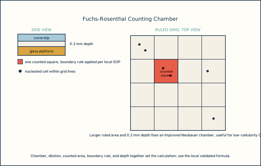
Manual CSF cell counts are commonly performed with a Fuchs-Rosenthal chamber, which provides a larger ruled counting area than an Improved Neubauer chamber and is useful for low-cellularity fluids like CSF because more volume is examined per count. The chamber type, coverslip seating, dilution (including whether the specimen is counted undiluted), counted area, boundary rule for cells touching a grid line, and chamber depth together determine the calculation. The laboratory applies its own validated, chamber-specific formula rather than a memorized multiplier, because those variables differ by chamber and by local procedure.
Certified pipettes or a validated commercial dilution system are used whenever a body fluid sample is diluted for counting. A pipette that delivers too much or too little volume creates a systematic dilution error, biasing every calculated result high or low even when duplicate chamber sides agree. Before use, the chamber grid lines must be bright and unobscured: scratches, dirt, or debris can hide or mimic a boundary, so different readers may include different cells and produce nonrepeatable counts. For manual body fluid cell counts the laboratory analyzes at least one cell count control in duplicate, or uses a procedural control such as correlation with a stained smear, according to its validated procedure.
A count chamber measures nucleated cells and red cells in a defined volume. It is not a diagnosis and not a definitive cellular differential; morphologic classification of the cells present is a separate step. Where unstained bright-field microscopy cannot reliably distinguish erythrocytes from other cells, the laboratory has an additional confirmation step available, such as an acid rinse to lyse red cells or a supravital stain.
Automated hematology analyzers with a body-fluid mode can also perform CSF counts, but automated and manual counts are not inherently interchangeable, particularly at low cell concentrations or when abnormal cells or debris are present. In one lumbar-CSF comparison, an automated analyzer's sensitivity against a manual reference threshold of 6 cells/uL was reported at 100% with specificity 75%, while both reached 100% at a 20 cells/uL threshold; those numbers describe that study's comparison thresholds and analyzer, not a general reference limit. In a separate drainage-fluid comparison, automated results ran higher than manual counts with proportional differences, so a laboratory tracking results over time needs to know which method produced each value.
If a fluid specimen is partially clotted or shows cell clumps or debris on the counting chamber, the laboratory indicates in the report that the result may be inaccurate. A clotted CSF specimen can trap cells unevenly and make a manual count nonrepresentative; the finding is examined and reported as a limitation under local rejection or qualification policy rather than silently reported as a normal count.
The chamber and the duplicate-side check are the quality control for a manual CSF count, so a flagged disagreement between sides is a stop-and-investigate signal, not a number to average away.
Illustrative drawing — this picture was drawn rather than captured.
Knowledge checks
Reading and checks are open. Sign in only to save.
Knowledge check 1
Knowledge check 2
Knowledge check 3
Knowledge check 4
Section status
Finish this section
Reading and checks are open. Sign in only to save.
The module finishes after every required section is marked done and every check in those sections is correct.