Required section · Section 3 of 6
Counting, viscosity, and reading the compensator
Gross viscosity in synovial fluid reflects hyaluronate polymerization, but protein content, cell numbers, temperature, and enzyme activity all affect it too, so viscosity is not a universal disease cutoff. When viscosity interferes with pipetting or counting, hyaluronidase can be added to reduce it before analysis, but that step introduces a dilution that must be calculated and it is only appropriate within a validated procedure; it is not a routine shortcut applied to every specimen.
Manual and automated body fluid counts both require method-specific validation covering specimen type, dilution scheme, reportable range, flags, and interferences. A high-cell specimen may need a measured dilution before counting, and the reported dilution correction is only valid within the laboratory's own validated procedure. The College of American Pathologists (CAP) Hematology, Coagulation, and Clinical Microscopy checklist requires that manual body fluid cell counts be performed in duplicate, with defined limits of agreement between the replicate counts and corrective action when those limits are not met.
A direct wet preparation lets you search native fluid for crystals without the artifact a stain can introduce. A stained cytocentrifuge (cytospin) preparation adds cellular morphologic detail and can concentrate a low-cellularity specimen, improving detection of both cells and particulate crystal material. CAP guidance favors stained cytocentrifuge preparations over unstained hemocytometer differentials for classifying body fluid nucleated cell types, reserving hemocytometer-based differentials for narrow uses such as distinguishing polymorphonuclear from mononuclear cells. When the counting method cannot reliably separate white blood cells from other nucleated cells, the count is reported as total nucleated cells (TNC), not as a white blood cell count, and the percentage differential from the stained slide covers every nucleated cell type observed, not only leukocytes.
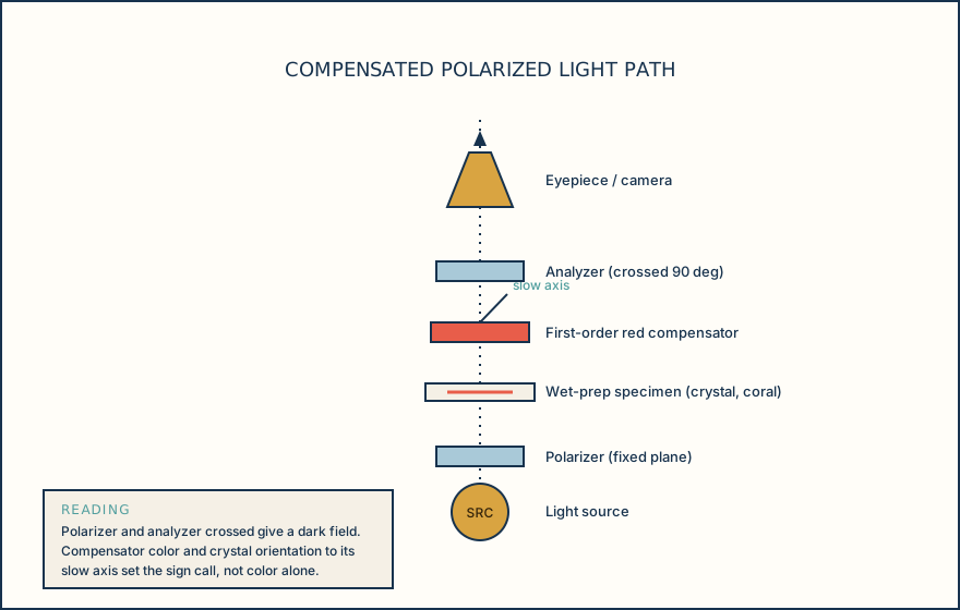
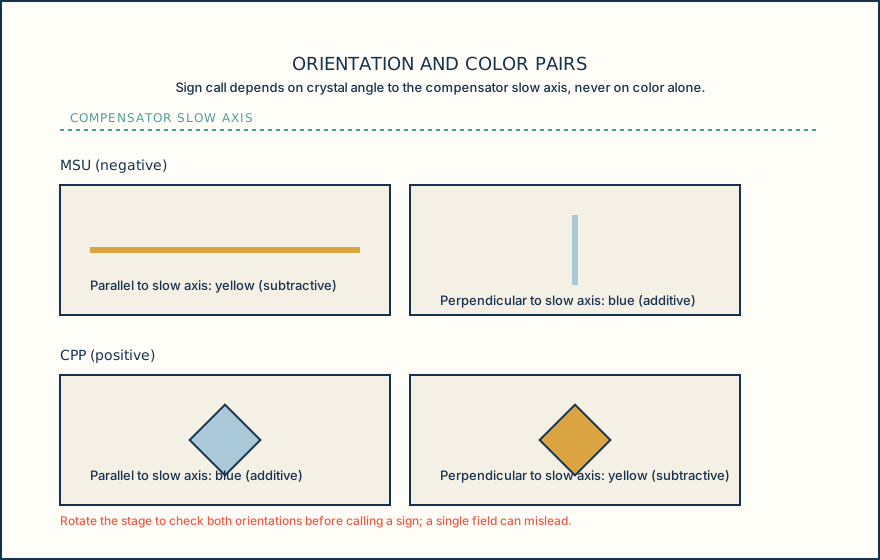
Compensated polarized light microscopy adds a polarizer and an analyzer set at 90 degrees to create a dark field, plus a first-order red compensator with a marked slow axis that lets you read a crystal's birefringence sign from its color and orientation. MSU crystals are usually slender needles with strong negative birefringence: with the standard compensator convention they appear yellow when the long axis is parallel to the slow axis and blue when perpendicular. CPP crystals are commonly rhomboid, rod-like, or parallelepiped shapes with weak positive birefringence, showing the opposite pairing, blue when parallel and yellow when perpendicular to the slow axis; CPP is smaller and less birefringent than MSU overall, and some CPP crystals show no birefringence at all, so a negative or equivocal field still needs cautious wording rather than a flat negative call.
CAP's crystal morphology requirement calls for scanning first at low power, commonly 10x, to locate candidate structures, then confirming shape and birefringence at higher power such as 40x or 100x, while distinguishing dust, scratches, and slide debris from genuine crystals. A defined local procedure for crystal identification and for who reviews a positive or ambiguous finding is part of that same expectation.
Read the compensator color together with the crystal's orientation on every candidate field, because color alone, without a documented orientation relative to the slow axis, does not establish a birefringence sign.
Illustrative drawing — this picture was drawn rather than captured.
Illustrative drawing — this picture was drawn rather than captured.
Knowledge checks
Reading and checks are open. Sign in only to save.
Knowledge check 1
Knowledge check 2
Section status
Finish this section
Reading and checks are open. Sign in only to save.
The module finishes after every required section is marked done and every check in those sections is correct.