Required section · Section 5 of 6
Your call at the microscope
You are handed the same case fluid and asked to work through three decisions: whether the specimen aliquots are suitable for the tests ordered, how to characterize what you see under compensated polarized light, and what to tell the ordering clinician about what the crystal finding does and does not mean. Each decision uses evidence you already have on the case report; none of them require inventing a new cutoff.
Specimen suitability comes first. A short-filled EDTA tube, a native crystal aliquot, and a protected sterile culture aliquot are all present, and none of the tubes shows a clot. The question is not whether the specimen is perfect; it is whether each aliquot is fit for the specific test it will support, and what limitation, if any, has to travel with the result.
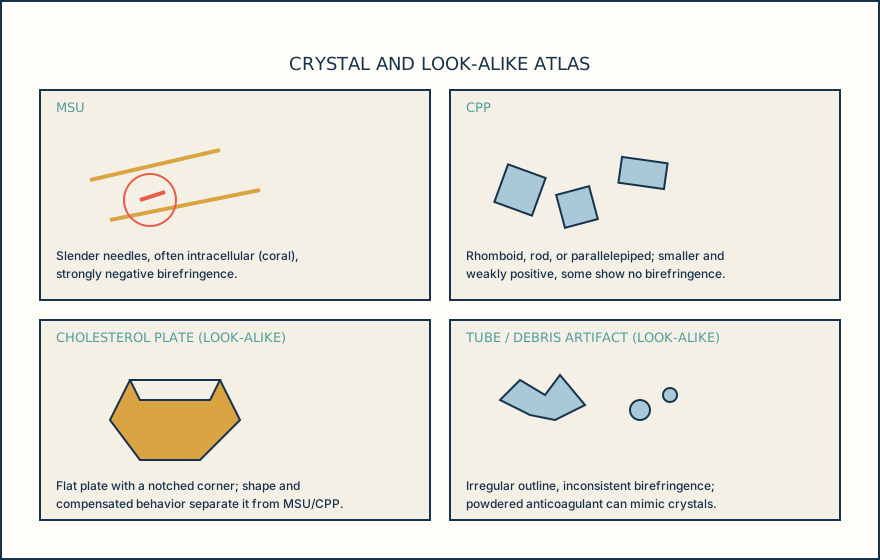
Crystal characterization is the second decision. Shape (needle versus rhomboid), birefringence sign (negative versus positive), the color pairing at two orientations to the compensator slow axis, and intracellular location together build the identification. Basic calcium phosphate crystals sit below what compensated polarized light can resolve and are nonbirefringent, so a negative compensated polarized light field never proves their absence; it only means MSU and CPP were not seen in that field.
Communication is the third decision. A synovial leukocyte count, including a count near or above 50,000 cells per microliter, does not reliably separate infection from crystal-induced inflammation once crystals are already present in the fluid, because crystal-positive fluid can also be culture-positive. The learner decision below asks you to reject the claim that a positive crystal finding rules out septic arthritis, and to choose the action that keeps the infection workup open.
A crystal identification and an infection workup are two separate lines of evidence that both belong on the same report until the culture result is final.
Illustrative drawing — this picture was drawn rather than captured.
Ordering exercise
Put this case's crystal examination workflow in the order that supports a defensible identification, from aliquot receipt to the final report note.
1. Switch to high power and apply the compensator
Confirm shape and read color at two orientations to the compensator slow axis to establish birefringence sign.
2. Report the crystal finding with the infection caveat
Release the identification while keeping the Gram stain and culture workup open rather than closed by the crystal result.
3. Confirm the crystal aliquot is native, additive-free fluid
Verify the tube intended for crystal search carries no anticoagulant that could generate look-alike material.
4. Note intracellular versus extracellular location
Record whether crystals are seen inside neutrophils, which strengthens but does not replace the morphology and sign findings.
5. Scan the wet preparation at low power
Search broadly at 10x to locate candidate needle- or rhomboid-shaped structures before committing to high power.
Knowledge checks
Reading and checks are open. Sign in only to save.
Knowledge check 1
Knowledge check 2
Knowledge check 3
Section status
Finish this section
Reading and checks are open. Sign in only to save.
The module finishes after every required section is marked done and every check in those sections is correct.