Required section · Section 2 of 6
The Cell-Based Model: What Actually Happens
The modern working model of hemostasis, described by Hoffman and Monroe, replaces the old linear intrinsic/extrinsic cascade with three overlapping cell-surface phases: initiation, amplification, and propagation. These phases are not a strict sequence with a clean handoff between steps. They overlap in time and in place, and none of them corresponds to a step you can point to in a citrate tube on an analyzer.
Initiation happens on a tissue-factor-bearing cell. Tissue factor complexed with factor VIIa activates small amounts of factor Xa and generates a trace of thrombin. That trace thrombin is far too little to clot blood by itself, but it is enough to reach nearby platelets.
Amplification is what that trace thrombin does next: it activates platelets and the cofactors V, VIII, and XI, building an activated platelet surface primed for large-scale enzyme assembly. Propagation follows on that platelet surface, where intrinsic tenase and prothrombinase complexes drive a large thrombin burst, far larger than anything generated during initiation.
That thrombin burst cleaves fibrinogen into fibrin monomers, which polymerize into a fibrin network. Thrombin and calcium activate factor XIII, and factor XIIIa then covalently cross-links that fibrin to stabilize the clot. In parallel, plasminogen activation on fibrin generates plasmin, while antifibrinolytic mechanisms restrain how fast that fibrin is broken down, so clot formation and clot breakdown are held in balance rather than running unopposed.
Illustrative drawing — this picture was drawn rather than captured.
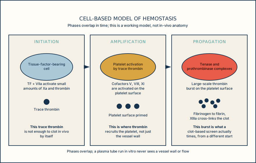
The overlapping phases of the cell-based model, from a tissue-factor-bearing cell to a stabilized, balanced fibrin clot.
Initiation
Tissue factor and factor VIIa on a tissue-factor-bearing cell activate limited factor Xa and generate trace thrombin.
Amplification
Trace thrombin activates platelets and cofactors V, VIII, and XI, priming the platelet surface.
Propagation
Tenase and prothrombinase complexes on the platelet surface drive a large-scale thrombin burst.
Fibrin formation and stabilization
Thrombin converts fibrinogen to fibrin monomers that polymerize, and factor XIIIa cross-links the network.
Fibrinolytic balance
Plasminogen activation on fibrin generates plasmin while antifibrinolytic mechanisms restrain breakdown, keeping formation and lysis balanced.
Knowledge checks
Reading and checks are open. Sign in only to save.
Knowledge check 1
Knowledge check 2
Section status
Finish this section
Reading and checks are open. Sign in only to save.
The module finishes after every required section is marked done and every check in those sections is correct.