Required section · Section 5 of 6
Reading the Pattern and Checking the Specimen
A screening pattern is only as good as the specimen behind it. Before you interpret any prolonged clot-based result, the same question always comes first: was this a citrate tube the laboratory can trust? Plasma-based clotting screens require citrated, platelet-poor plasma, and they are sensitive to collection, transport, and processing problems in ways many other tests are not.
The conventional collection system is buffered 3.2% citrate at a 9:1 whole-blood-to-citrate ratio. An underfilled tube leaves excess citrate relative to the plasma volume actually present, and that excess citrate can prolong clot-based results on its own, with no factor problem involved at all. A patient with an elevated hematocrit needs a locally validated citrate adjustment for the same reason: fixed citrate becomes excessive once there is less plasma per tube to begin with.
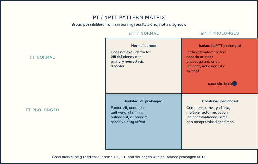
In the guided case, the tube was filled to the line, gently mixed, and was not hemolyzed, icteric, lipemic, or clotted, so this specific isolated aPTT prolongation is not explained by an obvious specimen problem. That does not make specimen review optional; it makes it the first check you complete and document, every time, before the pattern itself gets your attention.
Use the pattern matrix below to place a result before you go further. An isolated PT prolongation points broadly toward factor VII, a common-pathway effect, a vitamin K antagonist, or a reagent-sensitive drug effect. An isolated aPTT prolongation, like this case, points broadly toward intrinsic or contact-pathway factors, a heparin or other anticoagulant effect, or an inhibitor. Combined PT and aPTT prolongation can reflect a common-pathway effect, multiple factor reduction, an inhibitor or anticoagulant, or a compromised specimen. None of these four cells hands you a diagnosis; each one hands you a short, defensible list of what to check next.
Confirm the specimen is trustworthy first, place the pattern in the matrix second, and only then decide what escalation the pattern earns.
Illustrative drawing — this picture was drawn rather than captured.
| Specimen issue | Expected effect on clot-based screen | Laboratory action |
|---|---|---|
| Underfilled citrate tube | Excess citrate relative to plasma can prolong clot-based results | Assess against local rejection/recollection criteria before testing |
| Elevated hematocrit, no citrate adjustment | Fixed citrate becomes excessive relative to available plasma | Apply locally validated citrate adjustment per policy |
| Clotted specimen | Consumed factors and platelets give unreliable results | Reject and request recollection per policy |
| Grossly hemolyzed or lipemic specimen | Can interfere with clot detection depending on method | Assess against local interference policy before release |
Ordering exercise
Put the bench workflow for a citrate coagulation specimen in the order it should actually happen, from receipt to result release.
1. Check timing and transport
Compare draw, processing, and testing times against the method's stability limits.
2. Release the result
Report the result only once specimen quality has been confirmed acceptable.
3. Inspect the tube
Check for visible clots, hemolysis, and whether the fill line was reached.
4. Apply rejection or recollection policy
Reject or flag the specimen per local policy if any quality issue was found.
5. Confirm fill and hematocrit
Compare fill volume against the manufacturer line and flag elevated hematocrit for citrate adjustment.
Knowledge checks
Reading and checks are open. Sign in only to save.
Knowledge check 1
Knowledge check 2
Section status
Finish this section
Reading and checks are open. Sign in only to save.
The module finishes after every required section is marked done and every check in those sections is correct.