Required section · Section 4 of 6
Working the line-contamination case to a defensible conclusion
Return to the panel from the opening problem: citrate tube drawn 09:10 from a heparin-locked central line, PT 12.1 s (10.0-13.0), INR 1.0 (0.9-1.1), aPTT 118 s (25-35, high), TT greater than 180 s (14-21, high, no clot endpoint), heparin-calibrated anti-Xa 1.34 IU/mL (0.00-0.10, high). No anticoagulant history was submitted with the order, and the specimen itself is technically acceptable: not hemolyzed, not clotted, adequately filled.
Work the pattern against the mental model. PT and INR are normal, which is consistent with a heparin-type effect (heparin does not reliably affect PT the way it affects aPTT) and is not consistent with a VKA effect large enough to explain the aPTT. aPTT, TT, and heparin-calibrated anti-Xa are all markedly abnormal in the direction heparin produces. The magnitude of a heparin-calibrated anti-Xa result does not by itself rule a DOAC in or out, because a heparin-calibrated assay is not built to report DOAC concentration; the collection route is what narrows the pattern to heparin here. The specimen was drawn from a line documented as heparin locked, with no anticoagulant history submitted at all, so there is no DOAC exposure on record to weigh against the heparin-locked draw.
The finding that matters next is not the drug class, it is the source. The line is documented as heparin locked, and no infusion, dose, or timing was submitted, and no note confirms a validated line-draw procedure was followed. A heparin-exposed line can contaminate a coagulation specimen and produce exactly this picture: marked prolongation of heparin-sensitive clot assays and a falsely elevated anti-Xa, without reflecting the patient's actual circulating heparin exposure. The normal PT/INR does not rescue the interpretation, and it does not identify or rule out a drug; it only narrows which mechanisms are plausible.
The defensible action is to hold an anticoagulant-monitoring interpretation, communicate the collection concern, and obtain a repeat specimen by the locally approved peripheral collection process if testing is still clinically needed, rather than releasing the line-draw result as the patient's heparin status. A repeat peripheral specimen collected at 09:38, twenty-eight minutes later, returns PT 12.0 s, INR 1.0, aPTT 31 s, TT 18 s, and anti-Xa less than 0.10 IU/mL, every value inside the local reference band. See the paired chart below.
The paired pattern supports line contamination of the first specimen rather than a true systemic heparin effect, and it supports it because every abnormal marker normalized on a same-patient peripheral redraw within half an hour with no intervening treatment documented. This is a specimen-integrity finding, reported and resolved as one, not a dose-adjustment result.
Illustrative drawing — this picture was drawn rather than captured.
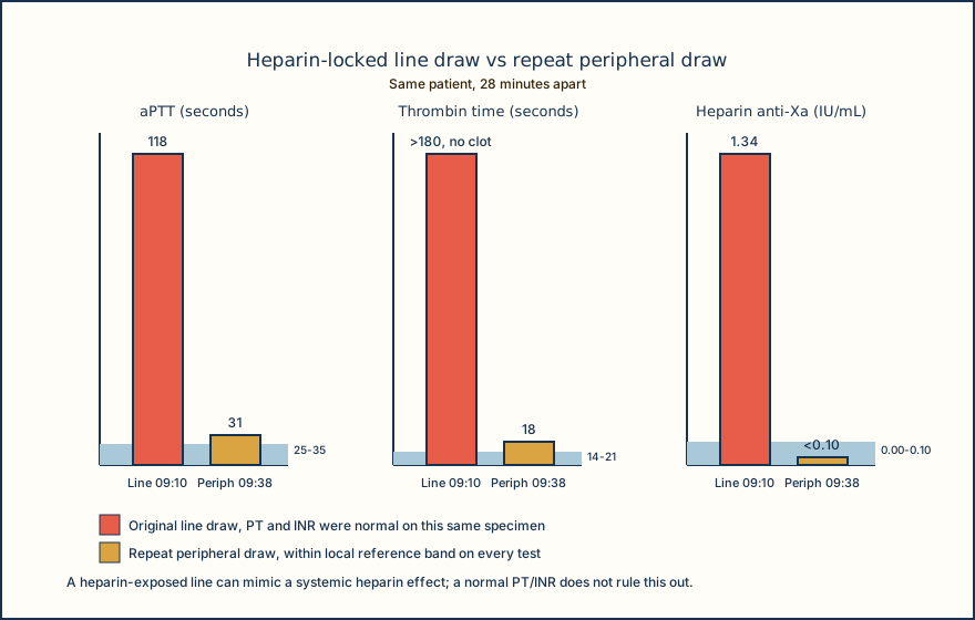
| Test | Local reference interval | Line draw 09:10 | Peripheral draw 09:38 |
|---|---|---|---|
| PT | 10.0-13.0 s | 12.1 s | 12.0 s |
| INR | 0.9-1.1 | 1.0 | 1.0 |
| aPTT | 25-35 s | 118 s, high | 31 s |
| TT | 14-21 s | >180 s, no clot endpoint | 18 s |
| Heparin-calibrated anti-Xa | 0.00-0.10 IU/mL | 1.34 IU/mL, high | <0.10 IU/mL |
Knowledge checks
Reading and checks are open. Sign in only to save.
Knowledge check 1
Knowledge check 2
Section status
Finish this section
Reading and checks are open. Sign in only to save.
The module finishes after every required section is marked done and every check in those sections is correct.