Required section · Section 3 of 6
Focused instruction: separate preparation effects from cell findings
Review the monolayer just behind the feathered edge for red-cell central pallor and distribution. Hypochromia is reduced hemoglobinization seen as central pallor clearly exceeding about one-third of RBC diameter. Mean corpuscular hemoglobin (MCH) is the calculated average hemoglobin mass per RBC, and mean corpuscular hemoglobin concentration (MCHC) is the calculated average hemoglobin concentration in packed RBC volume; use these indices rather than visual grading alone when available.
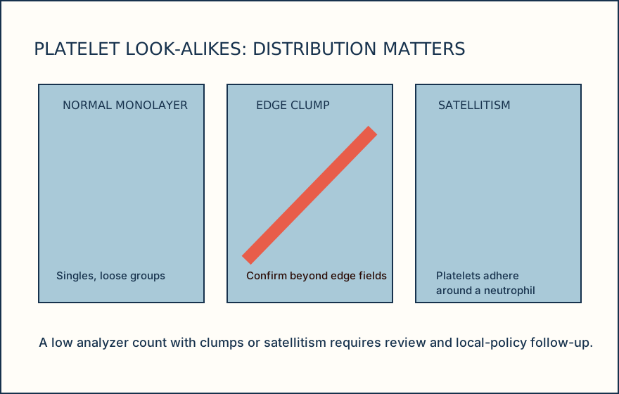
Normal platelets are spread as singles and small loose groups across the monolayer. Clumps at an edge or platelets rosetting around neutrophils, called satellitism, can make an automated count falsely low in an EDTA specimen. Giant platelets may also be misrepresented by impedance counting. A smear platelet estimate corroborates plausibility; it is not a replacement count.
Mean platelet volume (MPV) is an analyzer-estimated platelet size, not a direct microscopic measurement, and is method-dependent. Impedance methods estimate platelet volume from pulse amplitude, while optical methods derive size from light-scatter signal, so results are not numerically interchangeable. EDTA storage can change platelet shape and MPV over time.
Illustrative drawing — this picture was drawn rather than captured.
Ordering exercise
Place these actions in the order that supports a platelet-result discrepancy review.
1. Follow local policy
Document and escalate or obtain a citrate redraw when the procedure requires it.
2. Inspect edges
Search feathered and lateral edges for clumps or displaced material.
3. Compare analyzer result
Decide whether visible findings plausibly explain the automated result.
4. Inspect monolayer
Assess platelet distribution and RBC background where cells barely touch.
Knowledge checks
Reading and checks are open. Sign in only to save.
Knowledge check 1
Knowledge check 2
Section status
Finish this section
Reading and checks are open. Sign in only to save.
The module finishes after every required section is marked done and every check in those sections is correct.