Required section · Section 1 of 6
Opening laboratory problem: is this background interpretable?
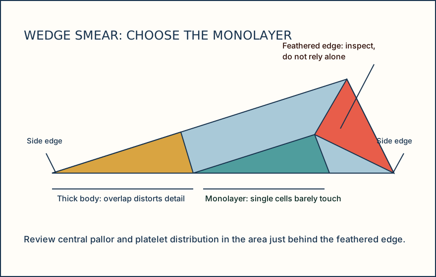
A peripheral smear accompanies an automated platelet count of 238 × 10^9/L with no analyzer flag. Before deciding that the result is ordinary, select the field that can support a description of red cells and platelets. The thick body, lateral edges, and feathered edge can each change what appears abundant or distorted. The question is whether the normal background is visible before any abnormality is named.
A manual wedge smear from well-mixed K2/K3-EDTA whole blood is stained with Wright or Wright-Giemsa stain. A single ideal field does not establish disease; the method shows what a well-prepared monolayer looks like and which preparation findings can mislead that view.
Start by looking at distribution rather than color alone. A usable field has red cells largely single layered and barely touching, with platelets available to inspect across the field. A concentrated edge finding is evidence to investigate, not a patient conclusion. Choose the field before you choose the morphology term.
Illustrative drawing — this picture was drawn rather than captured.
Knowledge checks
Reading and checks are open. Sign in only to save.
Knowledge check 1
Section status
Finish this section
Reading and checks are open. Sign in only to save.
The module finishes after every required section is marked done and every check in those sections is correct.