Required section · Section 3 of 6
Patterns that point to an interference
Cold-reactive IgM can agglutinate red cells below body temperature. On a method-specific histogram this can produce an irregular, broadened, high-volume aggregate pattern rather than a reliable cell-volume distribution. Aggregates may be counted as fewer, larger particles: RBC count and hematocrit fall while MCV, MCH, and especially MCHC rise; lysed-sample hemoglobin is comparatively reliable. Confirm with visible RBC agglutination on the original smear and apply a locally validated warmed rerun or warm recollection pathway.
Lipemic plasma is a turbidity-related photometric hemoglobin bias: inspect for lipemia, review the hemoglobin channel and internal relationships, and use a validated alternate hemoglobin approach or hold/escalate if it remains unresolved. Cell-free hemoglobin can also raise the photometric hemoglobin result, but requires separate evidence of free hemoglobin and a specimen/context review; a plasma observation alone does not establish its source. RBC destruction can occur in vitro or in vivo and is a separate process that can reduce intact RBC count and hematocrit; inspect specimen appearance, smear, collection history, and clinical context, then recollect or escalate when the destruction pathway cannot be resolved.
EDTA-dependent clumping makes the platelet count unreliable but does not exclude true thrombocytopenia. Confirm clumps on the original smear. Giant platelets can be undercounted above an impedance gate, while fragments or microcytes can be overcounted inside it; use direct morphology and an available validated alternate method to determine the reportable result.
For a short-filled K2EDTA tube, first confirm fill and specimen integrity, then review for relative anticoagulant excess (including reduced MCV, increased MCHC, and distorted morphology), inspect the original smear, and obtain a suitable recollection when the locally validated reportability limit is not met. For extreme leukocytosis, review analyzer linearity, flags, platelet separation, specimen, and original smear; use the validated dilution or alternate method, determine whether the analyzer or laboratory information system has already corrected the diluted rerun, compare evidence, then release only a verified result or suppress, recollect, consult, or escalate.
Illustrative drawing — this picture was drawn rather than captured.
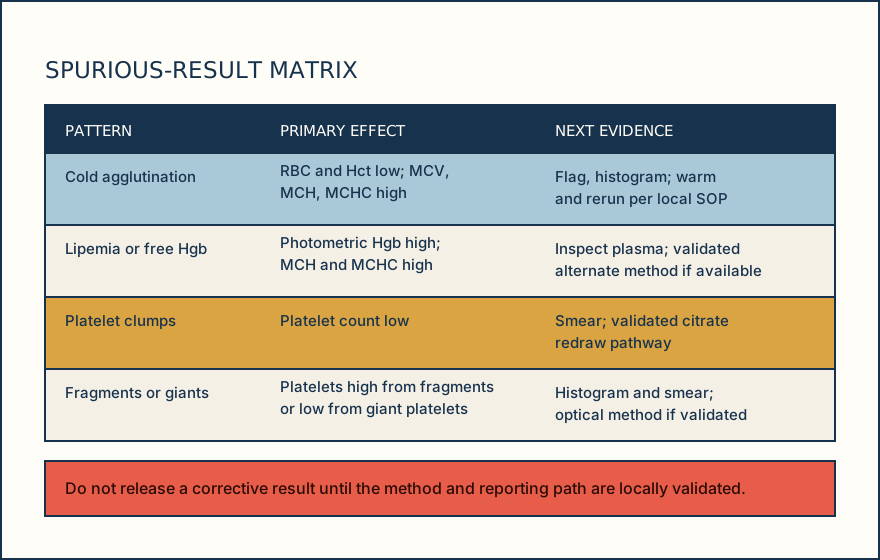
| Interference | Likely distorted result | Useful evidence | Bounded action |
|---|---|---|---|
| Cold agglutination | RBC and Hct low; MCV, MCH, MCHC high | Irregular/broad high-volume aggregate pattern and RBC clumps on original smear | Validated warming/rerun; if unresolved, warm recollection or consultation |
| Lipemic photometric bias | Hgb, MCH, MCHC high | Turbid plasma and channel/consistency review | Validated alternate Hgb approach; compare relationships before release |
| Cell-free hemoglobin | Photometric Hgb, MCH, MCHC may be high | Free-hemoglobin evidence plus specimen and clinical-context review | Identify source where possible; hold, recollect, or escalate if unresolved |
| RBC destruction | Intact RBC/Hct may be low; photometric Hgb interpretation may be affected | Specimen appearance, original smear, collection history, and clinical context | Distinguish in vitro from clinical context; recollect or escalate when unresolved |
| EDTA platelet clumps | Platelets unreliable, often low | Clump flag and clump-positive original smear | Hold; use valid alternate-anticoagulant path only after clump-free citrate smear |
| Giant platelets / fragments / microcytes | Platelets low with giants, high with fragments or microcytes | Histogram/gate review and labeled morphology | Validated optical/fluorescent method or manual review; escalate unresolved discordance |
| Short-filled K2EDTA | MCV may be low and MCHC high; morphology may be distorted | Fill assessment, analyzer relationships, and original smear | Use local reportability limit; recollect a suitable specimen or suppress/escalate |
| Extreme leukocytosis | WBC may exceed linearity; platelet separation may be compromised | Linearity/flag review, specimen, original smear, and platelet consistency | Validated dilution or alternate method; check for prior system correction, verify, then release or suppress/escalate |
Knowledge checks
Reading and checks are open. Sign in only to save.
Knowledge check 1
Knowledge check 2
Section status
Finish this section
Reading and checks are open. Sign in only to save.
The module finishes after every required section is marked done and every check in those sections is correct.