Required section · Section 4 of 6
Work the dimorphic case
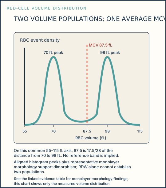
The MCV of 87.5 fL is an average. Peaks near 70 fL and 98 fL are proportional positions on the same volume axis, but the histogram alone does not establish dimorphism. The representative monolayer supplies the required morphology evidence: microcytic hypochromic cells and a discrete normocytic population are both present. Together, the film and analyzer evidence support a dimorphic population rather than continuous anisocytosis.
The low MCHC of 31.5 g/dL supports hypochromia somewhere in the aggregate red-cell population; it cannot assign hypochromia to the microcytes specifically. RDW-CV of 19.8% supports increased volume variation but cannot establish two populations. Polychromasia is concordant with the 2.8% reticulocyte percentage, but neither establishes the absolute marrow response without ARC and local convention.
Recent transfusion, treatment effect, combined nutritional deficiencies, and sideroblastic processes remain possible explanations for dimorphism. The morphology cannot establish which applies. When film and analyzer agree, report approved dimorphic morphology. When they disagree, follow the interference path: review specimen, flags, and original film; apply a validated verification step; then release, recollect, suppress, or escalate according to evidence.
Illustrative drawing — this picture was drawn rather than captured.
| Evidence | What it measures or shows | Bounded interpretation |
|---|---|---|
| MCV 87.5 fL | Average cell volume | Does not exclude two size populations |
| RDW-CV 19.8% | Variation in red-cell volume | Supports increased variation; correlate with smear |
| Histogram peaks 70 and 98 fL; MCV 87.5 fL | Measured volume distribution and calculated mean volume | Aligned peaks plus representative film support two populations; the mean does not establish uniformity |
| Microcytic hypochromic plus normocytic cells | Monolayer morphology | Supports increased variation but cannot establish dimorphism alone |
| Polychromasia and reticulocytes 2.8% | Stain observation and numeric result | Concordant only; ARC and local convention are needed to quantify response |
Knowledge checks
Reading and checks are open. Sign in only to save.
Knowledge check 1
Knowledge check 2
Section status
Finish this section
Reading and checks are open. Sign in only to save.
The module finishes after every required section is marked done and every check in those sections is correct.