Required section · Section 3 of 6
Make the indices accountable to method and morphology
ARC is primary; MCV and RDW refine the pattern. Under the local validated smear-review convention, ‘smear reviewed in optimal monolayer’ is recorded. Findings are stated positively only when actually observed, and a negative is stated only as a review finding—for example, ‘no schistocytes identified in 1,000 reviewed RBCs’—not as an analyzer-flag inference.
Each morphology described here is named with its defining observation: hypochromic microcytes are small RBCs with increased central pallor; macro-ovalocytes are large oval RBCs; hypersegmented neutrophils have more than five nuclear lobes; spherocytes are dense RBCs with reduced central pallor; polychromasia is diffusely bluish young RBC cytoplasm; and schistocytes are RBC fragments with sharp angles and straight borders. These are pattern evidence, not diagnoses.
Low ferritin and low transferrin saturation (TSAT) support restricted iron availability. After iron studies are already completed, do not label iron availability the next domain; identify the remaining bounded question or stop with an explicit missing-data outcome.
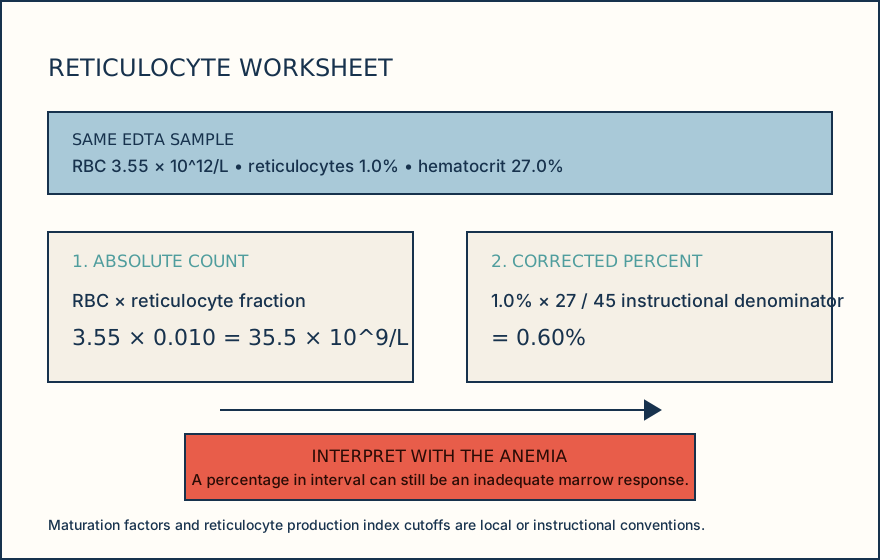
Illustrative drawing — this picture was drawn rather than captured.
Knowledge checks
Reading and checks are open. Sign in only to save.
Knowledge check 1
Section status
Finish this section
Reading and checks are open. Sign in only to save.
The module finishes after every required section is marked done and every check in those sections is correct.