Required section · Section 2 of 6
Build a reusable model of reactive change
Toxic granulation is increased prominence of primary, azurophilic granules in neutrophils. Its conspicuity varies with stain intensity and film quality, so compare cells across the film and with a validated local atlas. Döhle bodies are pale blue cytoplasmic inclusions that correspond ultrastructurally to rough endoplasmic reticulum aggregates.
Cytoplasmic vacuoles may accompany activation or phagocytosis, but they are not specific for infection or sepsis. Delayed preparation of a film from EDTA-anticoagulated blood can cause vacuolation, degranulation, and degenerative nuclear change. Collection-to-smear time and storage conditions are therefore part of the morphology observation.
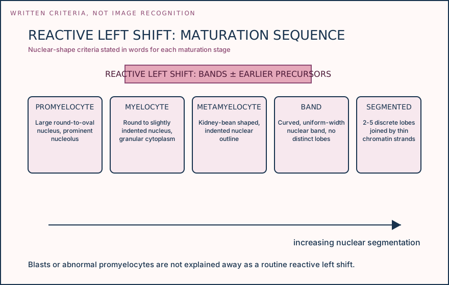
A reactive left shift means increased circulating granulocytic precursors, commonly bands with possible metamyelocytes and myelocytes. Blasts, abnormal promyelocytes, or an otherwise unexplained immature population require a different pathway rather than routine reactive labeling. Name each visible feature, then state what specimen and maturation context makes that feature interpretable.
Illustrative drawing — this picture was drawn rather than captured.
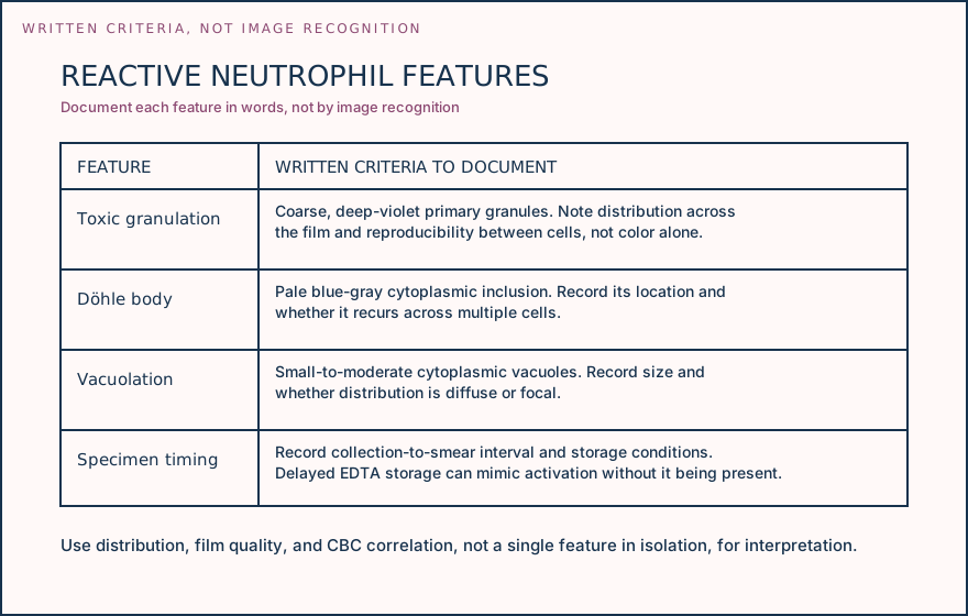
Illustrative drawing — this picture was drawn rather than captured.
Use this sequence before assigning a reactive morphology interpretation.
Observe and describe
Record granules, inclusions, vacuoles, nuclear detail, distribution, and focus behavior.
Add specimen and CBC context
Check collection-to-smear interval, storage, film quality, differential, and analyzer flags.
Classify the look-alike
Separate reactive features from stain artifact, storage degeneration, inherited morphology, and concerning immature cells.
Apply local policy
Release the approved comment, request a fresh smear when indicated, or escalate the review.
Knowledge checks
Reading and checks are open. Sign in only to save.
Knowledge check 1
Knowledge check 2
Section status
Finish this section
Reading and checks are open. Sign in only to save.
The module finishes after every required section is marked done and every check in those sections is correct.