Required section · Section 4 of 6
Guided cases: spurious and true low platelet counts
This case was collected in K2-EDTA at 09:18 and analyzed at 10:02 on an impedance platelet channel. Platelets are 34 × 10^9/L with platelet-clump and abnormal-distribution flags. Mean platelet volume is suppressed or not reported, and the histogram remains elevated at its high-volume boundary rather than returning cleanly to baseline. There is no visible clot in the tube. That boundary finding is nonspecific and cannot identify clumping without the smear.
The well-made smear has numerous platelet aggregates at the feathered and lateral edges. It has no fibrin strands and no schistocyte predominance. The aggregates make the 34 × 10^9/L result analytically unreliable; the instrument result cannot determine how many platelets are contained in aggregates or how much they changed the count. It is not reportable as a reliable platelet count.
At 10:25, a fresh EDTA specimen and properly filled alternate-anticoagulant specimen are collected under the local pseudothrombocytopenia procedure. The fresh EDTA smear again has edge clumps, while the alternate-tube smear has no clumps and the locally validated method returns 178 × 10^9/L. Release only the alternate result and comment authorized by that procedure, document EDTA-associated clumping, and do not transmit a verified critical call from the unreliable 34. In a clumped EDTA specimen, a confirmed alternate result can be released only when the local validated procedure authorizes it.
As a contrast case, a separate acceptable EDTA specimen has platelets of 42 × 10^9/L, no platelet-clump flag, and a histogram that returns cleanly at both boundaries. Its well-made smear shows reduced, evenly distributed individual platelets with no aggregates, fibrin, satellitism, RBC-fragment predominance, or dominant giant-platelet population. A locally validated optical result is concordant at 44 × 10^9/L. This evidence supports release of a verified true thrombocytopenic result and any urgent communication required by local policy; document the method and communication. It still does not establish the clinical cause.
Illustrative drawing — this picture was drawn rather than captured.
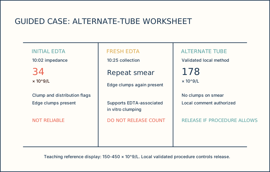
| Time | Specimen or method | Platelets | Key evidence | Disposition |
|---|---|---|---|---|
| 09:18 | K2-EDTA collection | Not yet analyzed | No visible clot | Proceed to CBC review |
| 10:02 | Impedance channel | 34 × 10^9/L | Clump and distribution flags; abnormal histogram | Do not release as reliable |
| 10:02 | EDTA smear | Not applicable | Numerous lateral and feathered-edge aggregates | Investigate EDTA-associated clumping |
| 10:25 | Fresh EDTA smear | Not applicable | Edge clumps again present | Do not release EDTA count |
| 10:25 | Alternate tube, validated method | 178 × 10^9/L | No clumps on smear | Release only if local procedure authorizes |
| Specimen or method | Platelets | Reliability evidence | Authorized disposition |
|---|---|---|---|
| Acceptable K2-EDTA specimen | 42 × 10^9/L by impedance | Correct tube and fill; no visible clot; no platelet-clump flag; histogram returns to baseline | Continue smear and method correlation |
| Well-made EDTA smear | Not applicable | Reduced, evenly distributed individual platelets; no aggregates, fibrin, satellitism, fragment predominance, or dominant giant-platelet population | Supports a credible platelet population |
| Locally validated optical method | 44 × 10^9/L | Concordant with the impedance result and suitable for the specimen | Release verified low result; communicate only as required by local policy and document method |
Knowledge checks
Reading and checks are open. Sign in only to save.
Knowledge check 1
Section status
Finish this section
Reading and checks are open. Sign in only to save.
The module finishes after every required section is marked done and every check in those sections is correct.