Required section · Section 3 of 6
Dysplasia, Mimics, and Classification Boundaries
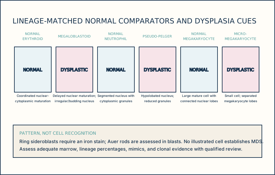
Dyserythropoiesis may include abnormal nuclear maturation, budding, irregular contours, multinuclearity, and megaloblastoid change (erythroid precursors with delayed nuclear maturation and relatively mature cytoplasm). Dysgranulopoiesis may include hypogranulation and hypolobation such as pseudo-Pelger forms (neutrophils with an abnormally bilobed or unsegmented nucleus). The term micromegakaryocytes means abnormally small megakaryocytes; a micromegakaryocyte is one such cell; separated megakaryocyte lobes are nuclear lobes with little or no connecting chromatin. These features support dysmegakaryopoiesis only when assessed as a reproducible lineage pattern. Ring sideroblasts are erythroid precursors with iron-laden mitochondria forming a perinuclear ring on an iron stain. Auer rods are needle-like azurophilic cytoplasmic inclusions in blasts and materially affect blast-category assessment.
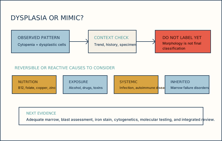
Before final interpretation, consider vitamin B12 or folate deficiency, copper deficiency, zinc excess, alcohol, drugs, toxins, infection, autoimmune disease, and inherited marrow disorders. Copper deficiency can cause anemia or neutropenia with vacuolated precursors, dysplasia, and ring sideroblasts. Correctable causes must be documented and addressed before a morphology pattern is used to classify a myeloid neoplasm.
WHO-HAEM5 calls the disease family myelodysplastic neoplasm, while ICC retains myelodysplastic syndrome in entity names. WHO groups MDS into defining-genetic-abnormality and morphologically defined entities and no longer uses single versus multilineage dysplasia as a subtype distinction. Both systems require a controlled classification procedure and qualified hematopathology review; the worksheet records observed findings and does not assign a subtype.
Illustrative drawing — this picture was drawn rather than captured.
Illustrative drawing — this picture was drawn rather than captured.
Knowledge checks
Reading and checks are open. Sign in only to save.
Knowledge check 1
Knowledge check 2
Section status
Finish this section
Reading and checks are open. Sign in only to save.
The module finishes after every required section is marked done and every check in those sections is correct.