Required section · Section 5 of 6
Your call: classify, correlate, and decide
Identify the cells you can identify, judge whether the preparation supports that identification, and decide what the pattern means when correlated with the other results. Separate confident calls from cells that need a second review.
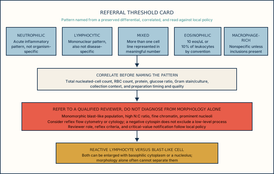
Refer a slide to a qualified reviewer when it shows a monomorphic population, a high nuclear-to-cytoplasmic ratio, fine chromatin, or prominent nucleoli. Reactive lymphocytes can partly mimic these features. A neutrophil-predominant inflammatory pattern alone, even at 62 percent, does not trigger referral. The laboratory sets reviewer qualifications, reflex flow cytometry or cytology criteria, and critical-value notification rules; do not rely on a universal numeric threshold.
Timing also matters for hemorrhage-related findings. RBCs, erythrophagocytosis, and siderophages do not all appear at once; they represent a sequence from fresh blood exposure through red-cell ingestion to iron breakdown. Use that sequence without assigning an exact hour, because the timing varies too much for any of these findings to serve as a precise clock.
Match the finding to the action it actually supports, correlate before you commit to a pattern name, and reserve referral for the specific features that call for it.
Illustrative drawing — this picture was drawn rather than captured.
Ordering exercise
A CSF specimen contains blood from an in vivo hemorrhage that occurred before the tap. Place these three findings in the sequence they are expected to appear over time, from earliest to latest.
1. Siderophages
Macrophages contain hemosiderin from breaking down ingested RBCs, supporting prior blood breakdown; onset and persistence vary too much to date the bleed precisely from this finding alone.
2. Intact RBCs with an otherwise unremarkable differential
Fresh blood is present in the CSF; at this point the RBCs are simply visible, not yet ingested or broken down by macrophages.
3. Erythrophagocytosis
Macrophages are seen with ingested intact or partially digested RBCs; this does not occur when blood and CSF are simply mixed outside the body, so it favors bleeding before collection.
Knowledge checks
Reading and checks are open. Sign in only to save.
Knowledge check 1
Knowledge check 2
Section status
Finish this section
Reading and checks are open. Sign in only to save.
The module finishes after every required section is marked done and every check in those sections is correct.