Required section · Section 3 of 6
Reading Adequacy, Contamination, and Pigment Before You Trust the Count
Squamous epithelial cells in a BAL specimen indicate contamination with upper-airway secretions picked up as the bronchoscope passed through the mouth and pharynx, not a distal airspace finding. A specimen with more than about 5% squamous cells is presumed contaminated with proximal material. Large numbers of ciliated bronchial epithelial cells point to the same problem from a different source: the lavage sampled proximal airway rather than distal alveolar spaces adequately. Neither finding is dangerous, but both undercut confidence in whatever the rest of the differential seems to show.
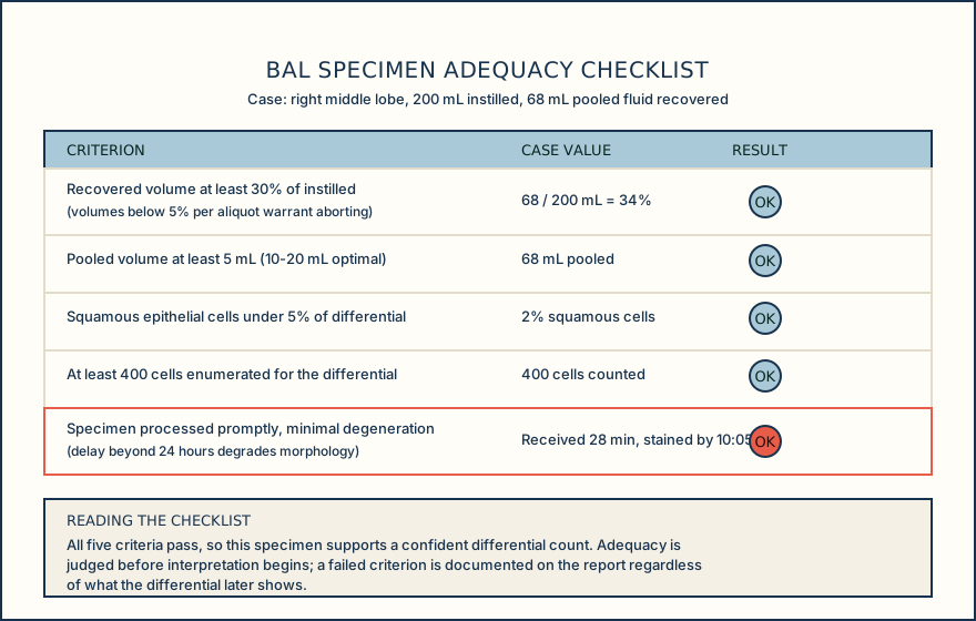
Other reasons a BAL specimen is considered inadequate include too few alveolar macrophages, as a rule of thumb under about 10 per high-power field, excess bronchial or squamous epithelial cells relative to macrophages, obscuring mucopurulent material, numerous red blood cells suggesting traumatic sampling rather than true hemorrhage, and significant cell degeneration. Degeneration reflects poor preservation from a transport delay, storage problem, or processing delay, and it can make cells difficult to classify with confidence even when the collection itself was reasonable.
Heavy mucus can physically obscure macrophages and lower confidence in the count; a grossly mucoid sample may need straining or treatment to loosen it before a slide is made. CAP's own smear-quality requirement expects uniform cell distribution, appropriate dilution so cells are not crowded, proper staining, adequate cell yield, and cell types that can be readily recognized, and expects a laboratory to maintain a reference file or atlas of body fluid cytomorphology at the bench to support that recognition.
Cytocentrifugation itself is not perfectly representative. It does not recover all cells with equal efficiency, and smaller cells, particularly lymphocytes, can be under-represented on the slide relative to their true proportion in the fluid. This is a known limitation of the preparation method, not a specimen defect, and it is one more reason a borderline differential should be read with some caution rather than treated as an exact count.
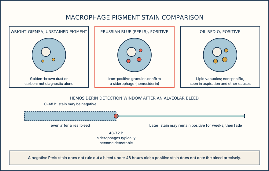
Alveolar macrophages carry information beyond their percentage. They are the largest mononuclear cell on the slide, with abundant cytoplasm that may contain phagocytosed material, dust, or pigment; heavy pigmentation is typical and expected in smokers. Hemosiderin-laden macrophages, or siderophages, are confirmed with a Prussian blue (Perls) iron stain, and they generally do not become detectable until roughly 48-72 hours after an alveolar bleeding event. A negative Perls stain therefore does not exclude a very recent hemorrhage under 48 hours old, and hemosiderin can also fade or clear well after a remote bleed, so the stain has a real timing blind spot in both directions.
Free, uningested red blood cells that increase across sequential aliquots are more supportive of active or recent bleeding than hemosiderin-laden macrophages alone, which reflect a somewhat older event. The Golde score, which grades roughly 100 macrophages 0-4 for staining intensity and sums the result, has been used in the literature as a semiquantitative severity index, but published cutoffs vary between studies and there is no single universal decision limit; any specific number used at the bench is a local or literature-sourced convention, not a fixed standard.
A lipid-laden macrophage index (LLMI), scoring Oil Red O staining of about 100 macrophages 0-4, has been used to support a clinical impression of aspiration or lipid-associated lung injury, but no single cutoff is universally accepted and a positive result does not by itself prove aspiration. Lipid-containing macrophages are nonspecific: they are also seen with infection, inflammation, smoking, pulmonary alveolar proteinosis, and other lipid exposures, so an LLMI result needs clinical and radiologic correlation before it means anything.
Read the specimen for contamination, mucus, degeneration, and recovered volume before the differential percentages are allowed to mean anything, and treat both the Golde score and the LLMI as literature-described indices with locally defined thresholds, not fixed cutoffs.
Illustrative drawing — this picture was drawn rather than captured.
Illustrative drawing — this picture was drawn rather than captured.
| Pattern | Threshold | Broad, nonspecific association |
|---|---|---|
| Lymphocytic | Lymphocytes above 15% | Sarcoidosis, hypersensitivity pneumonitis, drug reaction, other interstitial lung disease |
| Neutrophilic | Neutrophils above 3% | Acute lung injury, aspiration, suppurative infection |
| Eosinophilic | Eosinophils above 1% | Eosinophilic pneumonia in the appropriate clinical setting |
| Mast-cell | Mast cells above 0.5% | Reported in the literature; not detailed further here |
Knowledge checks
Reading and checks are open. Sign in only to save.
Knowledge check 1
Knowledge check 2
Section status
Finish this section
Reading and checks are open. Sign in only to save.
The module finishes after every required section is marked done and every check in those sections is correct.