Required section · Section 2 of 6
From Wedge to Slide: How a BAL Specimen Becomes a Differential
A BAL is collected by wedging the tip of a bronchoscope into a bronchopulmonary segment and instilling 100-300 mL of room-temperature saline in three to five sequential aliquots. Each aliquot is suctioned back before the next is instilled. Optimal sampling of the distal airspaces requires recovering at least 30% of the instilled volume overall; if an individual aliquot returns under 5%, the procedure is generally aborted because so little of what comes back will represent alveolar contents. The lobe and segment sampled should be documented on the requisition, because both the expected cellular composition and the reference range shift depending on where the lavage was taken.
Once fluid is back, the aliquots are pooled, and pooling all aliquots including the first is acceptable for a routine cellular differential. A minimum pooled volume of 5 mL is needed to run a cellular analysis at all; 10-20 mL is optimal. The specimen should reach the laboratory and be processed promptly. Waiting beyond about 24 hours, or storing the specimen improperly, degrades cell morphology and can make an otherwise adequate collection unreadable.
The differential itself is read from a stained cytocentrifuge (cytospin) preparation, most often Wright-Giemsa or May-Grunwald-Giemsa stain, rather than from an unstained hemocytometer count. Cytocentrifugation gives better morphologic detail and a higher rate of malignant-cell detection than unstained wet-preparation methods, which is why CAP's own requirement for body fluid cell differentials favors it. The method-defined target for a BAL differential is 400 nucleated cells, reported as a percentage of nucleated cells counted; that target is chosen to keep counting imprecision acceptable for cell types that make up only a small share of the differential, such as eosinophils or mast cells near their own pattern thresholds. When fewer than 400 cells can be enumerated because the specimen is hypocellular, a differential is still reported, but precision is worst for these minor populations: a percentage built on 100 cells carries far wider count-to-count variability than the same percentage built on 400, so a low-count differential should carry a comment on limited precision rather than being read with the same confidence as a full 400-cell count.
In a healthy nonsmoking adult, the expected BAL differential is alveolar macrophages above 85%, lymphocytes 10-15% with a CD4:CD8 ratio of roughly 0.9-2.5, neutrophils under 3%, eosinophils under 1%, and squamous or ciliated epithelial cells under 5%. Smoking status shifts this: smokers typically show a higher macrophage percentage and a correspondingly lower lymphocyte and neutrophil share. These numbers describe a single-center, pooled reference population, not a universal biological constant, and they set the baseline used throughout.
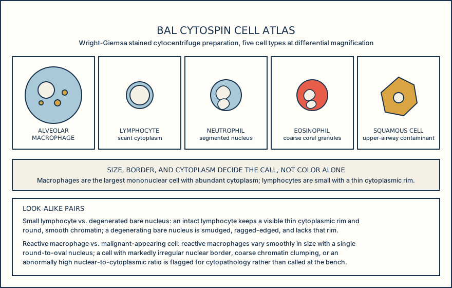
Cell identity is read from size, nuclear features, cell border, and cytoplasmic detail together, never from color or overall impression alone. Alveolar macrophages are the largest mononuclear cell on the slide, 15-80 micrometers, with a round-to-oval eccentric nucleus of fine chromatin and a small or inconspicuous nucleolus, an irregular ameboid cytoplasmic border, and abundant cytoplasm that is often foamy or vacuolated and may carry phagocytosed pigment, dust, or debris. Lymphocytes are small, 8-12 micrometers and only slightly larger than a red cell, with a round nucleus of dense, coarse chromatin filling most of the cell, a smooth round border, and a thin, scant, nongranular cytoplasmic rim. Neutrophils, 10-14 micrometers, have a nucleus segmented into two to five lobes joined by thin chromatin strands, a smooth round border, and pale, finely granular cytoplasm. Eosinophils, 12-17 micrometers, usually show a bilobed nucleus, a smooth round border, and cytoplasm packed with large, coarse, refractile orange-red granules. Squamous or ciliated bronchial epithelial cells are large, 30-60 micrometers for squamous forms, with a small, central-to-basal, often pyknotic nucleus; a polygonal, sharply defined border for squamous forms or a columnar border with a terminal bar for ciliated bronchial forms; and cytoplasm that is flat and keratinized (squamous) or columnar with visible surface cilia (bronchial). The same four features are what separate a genuine cell from the two categories of look-alike every BAL reader has to rule out: a degenerated bare nucleus, which matches the size of its parent cell's nucleus (most often lymphocyte-sized) but shows smudged, degenerating chromatin and no discernible cytoplasmic rim at all, the absence of any cytoplasm being what separates it from an intact lymphocyte; and a malignant-appearing cell, which is enlarged and variable from cell to cell (anisonucleosis), often larger than a reactive macrophage, with a markedly enlarged nucleus showing an irregular membrane, coarse or clumped chromatin, and a prominent or multiple nucleoli unlike a reactive macrophage's bland round nucleus, an irregular cell and nuclear border with a high nuclear-to-cytoplasmic ratio, and cytoplasm that is scant relative to nuclear size and sometimes abnormally vacuolated, a finding to refer rather than call at the bench.
Before reading a single cell, know what was collected, how it was pooled, and how it was stained, because each step shapes what the differential can and cannot tell you.
Illustrative drawing — this picture was drawn rather than captured.
| Cell type | Normal nonsmoking-adult range | Pattern threshold |
|---|---|---|
| Alveolar macrophages | Above 85% | Not applicable |
| Lymphocytes | 10-15% | Lymphocytic pattern above 15% |
| Neutrophils | Under 3% | Neutrophilic pattern above 3% |
| Eosinophils | Under 1% | Eosinophilic pattern above 1% |
| Mast cells | Under 0.5% | Mast-cell pattern above 0.5% |
From bronchoscopic wedge to a released BAL differential.
Wedge and instill
The bronchoscope is wedged in the target segment and 100-300 mL of saline is instilled in three to five sequential aliquots; the lobe and segment are recorded on the requisition.
Recover and pool
Each aliquot is suctioned back; at least 30% overall recovery is the adequacy target, and all aliquots including the first are pooled for a routine differential, with a minimum of 5 mL and an optimal 10-20 mL pooled volume.
Prepare the cytocentrifuge slide
The pooled fluid is processed promptly, generally within 24 hours, and a cytocentrifuge preparation is made and stained with Wright-Giemsa or May-Grunwald-Giemsa, the method favored over unstained hemocytometer preparations for classification and malignant-cell detection.
Count the differential
400 nucleated cells is the method-defined target for a BAL differential, enumerated on the stained slide and reported as a percentage of nucleated cells; a total nucleated cell count is reported alongside when the counting method cannot reliably separate white blood cells from other nucleated cells, and counts short of the target carry a precision comment, especially for minor cell populations.
Compare to the reference pattern
The differential is compared against the healthy nonsmoking adult range and the lymphocytic, neutrophilic, eosinophilic, and mast-cell pattern thresholds, producing a descriptive pattern label rather than a diagnosis.
Knowledge checks
Reading and checks are open. Sign in only to save.
Knowledge check 1
Section status
Finish this section
Reading and checks are open. Sign in only to save.
The module finishes after every required section is marked done and every check in those sections is correct.