Required section · Section 3 of 6
Why the simple pattern can fail
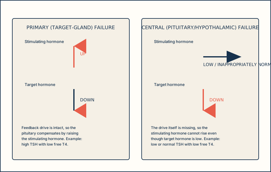
The primary-versus-central pattern above assumes a specimen drawn at a representative moment from a patient with no other physiologic or interference factor acting on the result. Five variables can break that assumption, and a bench scientist checks for them before treating an unexpected pattern as pathology rather than context.
Timing and pulsatility change what a single draw represents. ACTH and cortisol are pulsatile and circadian, peaking near waking and reaching a trough near midnight. A random cortisol draw at the wrong point in that rhythm can look abnormal without any disease present. TSH does not share that large-amplitude pulsatility, but it does have a modest nocturnal rise and is the more time-stable signal of the two axes covered here.
Total versus free hormone matters because most circulating cortisol and T4 travel bound to carrier proteins, and only the unbound, free fraction is biologically active. Estrogen, from pregnancy or oral contraceptives, raises thyroxine-binding globulin (TBG); total T4 rises with it while free T4 stays in its normal range because feedback holds the free fraction stable. A total-hormone assay and a free-hormone assay can therefore answer different questions from the same specimen, which is why free T4 rather than total T4 is preferred for hypothyroidism assessment outside pregnancy.
Acute and critical illness alter the axis by more than one mechanism at once. In non-thyroidal illness syndrome, sometimes called sick euthyroid pattern, T3 falls first, free T4 may fall as illness worsens, and TSH stays low, low-normal, or only mildly abnormal rather than rising the way primary hypothyroidism would produce. This pattern can look like central hypothyroidism on paper without any pituitary disease present, which is a real reason not to interpret thyroid testing acontextually during acute illness.
Reference intervals are population- and age-dependent. In one large U.S. population study, reported TSH upper limits rose from about 3.56 mIU/L at ages 20–29 to about 7.49 mIU/L at ages 80 years and older; these are study-specific illustrative examples, not this laboratory’s interval. A mildly elevated TSH in an older adult is not automatically evidence of primary hypothyroidism once age, assay, the local interval, and clinical context are considered. Assay interference is the fifth variable, covered in the learner-decision section.
Illustrative drawing — this picture was drawn rather than captured.
| Analyte | Result | This laboratory's reference interval | Age-adjusted context | Flag |
|---|---|---|---|---|
| TSH | 5.8 mIU/L | 0.40-4.50 mIU/L (general adult) | Reported upper limits rise with age; approx. 7.49 mIU/L at 80+ years in a large reference population | High by the general-adult interval |
| Free T4 | 0.9 ng/dL | 0.8-1.8 ng/dL | Within interval, low-normal | Not flagged |
| Patient context | 78 years old, outpatient, no acute illness or biotin history noted | - | - | - |
Knowledge checks
Reading and checks are open. Sign in only to save.
Knowledge check 1
Knowledge check 2
Knowledge check 3
Section status
Finish this section
Reading and checks are open. Sign in only to save.
The module finishes after every required section is marked done and every check in those sections is correct.