Required section · Section 2 of 6
Two layers, read separately
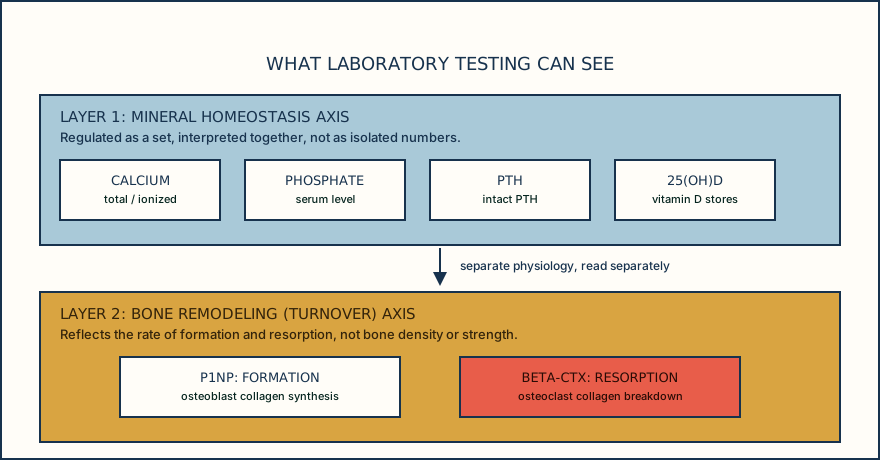
A bone-health laboratory panel has two layers, and mixing them up leads to wrong conclusions. The first layer is mineral homeostasis: calcium, phosphate, and parathyroid hormone (PTH) are regulated together and are read as a set, not as isolated numbers, with 25-hydroxyvitamin D (25(OH)D) as an input that affects calcium absorption. The second layer is bone remodeling: formation markers reflect osteoblast activity building new bone matrix, and resorption markers reflect osteoclast activity breaking existing bone down. These two layers answer different questions and use different tests.
A reasonable initial secondary-cause evaluation for low bone mass draws from the first layer plus a few general tests: complete blood count, calcium (with albumin or ionized calcium), phosphorus, alkaline phosphatase (ALP), creatinine or estimated glomerular filtration rate (eGFR), 25(OH)D, and often thyroid-stimulating hormone (TSH) and intact PTH. The goal is to find a treatable contributor to bone loss, such as hyperparathyroidism, renal impairment, or thyroid disease, before assuming the bone loss is primary.
Total ALP sits at the boundary between the two layers and is a good illustration of why source matters before interpretation. Total ALP is nonspecific: it rises from liver, bone, intestine, or placenta. An isolated elevation needs a source-narrowing step, such as gamma-glutamyl transferase (GGT) or bone-specific alkaline phosphatase (BSAP), to support or argue against a hepatobiliary or bone source, before it is read as a bone signal at all.
The diagram below lays out the workflow a laboratory scientist walks through when reading this panel: read the mineral axis as a set, screen for secondary causes, narrow an ambiguous ALP result, then layer in turnover markers if ordered. None of these steps, alone or together, diagnoses osteoporosis or estimates fracture risk; that determination uses DXA criteria or a recognized low-trauma fracture site meeting the cited diagnostic criteria. Know which layer a result belongs to before you decide what it means.
Illustrative drawing — this picture was drawn rather than captured.
Reading a bone-health laboratory panel, in order.
Read the mineral axis as a set
Look at calcium, phosphate, and PTH together, not one at a time; they are physiologically coupled and a normal-looking single value can still sit inside an abnormal pattern.
Screen for secondary contributors
Check renal function (creatinine/eGFR), thyroid status (TSH), and vitamin D stores (25(OH)D) for a treatable cause of bone loss before assuming the loss is primary.
Narrow an ambiguous ALP result
An isolated total ALP elevation is not automatically a bone signal: use GGT or BSAP to support or argue against a hepatobiliary or bone source before interpreting it as a bone signal.
Layer in turnover markers if ordered
P1NP (formation) and beta-CTX (resorption) add a remodeling-rate signal on top of the mineral axis, useful for monitoring, not for diagnosis.
Remember what none of this diagnoses
Osteoporosis diagnosis and fracture-risk estimation remain with DXA bone mineral density or a recognized low-trauma fracture site meeting the cited diagnostic criteria, not with any chemistry result.
Knowledge checks
Reading and checks are open. Sign in only to save.
Knowledge check 1
Knowledge check 2
Section status
Finish this section
Reading and checks are open. Sign in only to save.
The module finishes after every required section is marked done and every check in those sections is correct.