Required section · Section 3 of 6
Affinity shifts and method limits
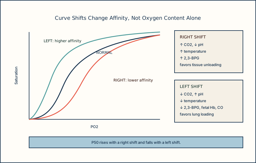
A right shift means lower hemoglobin oxygen affinity and a higher P50, favoring oxygen unloading in tissue. Increased CO2, decreased pH, increased temperature, and increased 2,3-BPG shift right. The Bohr effect describes reduced affinity when CO2 and hydrogen ion increase. A left shift means higher affinity and lower P50, favoring lung loading but potentially impairing unloading.
Decreased CO2, increased pH, decreased temperature, decreased 2,3-BPG, fetal hemoglobin, and carbon monoxide occupancy can shift left. 2,3-BPG is a red-cell glycolytic intermediate that preferentially binds deoxyhemoglobin and stabilizes the low-affinity state. Stored red cells lose 2,3-BPG, an initial blood-bank consideration after transfusion. Curve shifts explain affinity, not a substitute for checking hemoglobin concentration or measured fractions.
Carboxyhemoglobin and methemoglobin can make saturation conventions diverge. Functional sO2 excludes dyshemoglobins from its denominator, whereas FO2Hb includes them. Standard pulse oximetry cannot distinguish COHb from oxyhemoglobin and commonly trends toward about 85% with methemoglobinemia. Co-oximetry is required to quantify COHb and MetHb directly.
Air bubbles drive pO2 toward room air, liquid heparin can dilute or alter results, and delay permits cellular metabolism to lower pO2 and raise pCO2. A discordant result begins with specimen and method review, not a clinical treatment recommendation. Local validated procedure governs transport timing, rejection, reference intervals, result labels, competency, and comparability across instruments. When a saturation result conflicts with the picture, verify specimen integrity and request co-oximetry when fractions matter.
Illustrative drawing — this picture was drawn rather than captured.
| Condition | What changes | Pulse oximetry limit | Co-oximetry contribution |
|---|---|---|---|
| COHb | Carbon monoxide occupies hemoglobin | May read falsely normal to high | Quantifies COHb fraction |
| MetHb | Ferric hemoglobin cannot bind oxygen | Often trends toward about 85% | Quantifies MetHb fraction |
| No dyshemoglobin | Normal fractions expected | Estimates SpO2 under tested conditions | Measures O2Hb and HHb fractions |
Knowledge checks
Reading and checks are open. Sign in only to save.
Knowledge check 1
Knowledge check 2
Section status
Finish this section
Reading and checks are open. Sign in only to save.
The module finishes after every required section is marked done and every check in those sections is correct.