Required section · Section 3 of 6
Method details, agonists, and where each method breaks
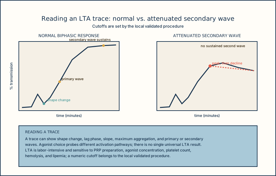
Light-transmission aggregometry (LTA) uses citrated platelet-rich plasma (PRP), stirred while a declared agonist is added, and measures increased light transmission as platelets aggregate and the suspension clears. A trace can show shape change, a lag phase, slope, maximum aggregation, and primary and secondary waves. Agonist selection matters: different agonists probe different activation pathways, so a panel of agonists, not one agonist, is what generates an interpretable pattern. LTA is labor-intensive and sensitive to PRP preparation, agonist concentration, platelet count, hemolysis, and lipemia, which is why it stays a reference method performed in laboratories with the staffing and volume to maintain it well.
Whole-blood impedance aggregometry uses citrated whole blood without a PRP preparation step. Activated platelets adhere to paired electrodes and recruit additional platelets, and the signal is a rise in electrical impedance, reported in ohms or as an instrument-derived area under the curve. Impedance aggregometry preserves red cells and white cells in the test system, which LTA does not, but it has its own platelet-count and hematocrit limitations, and it does not display the shape-change or reversibility information an LTA trace can show. An impedance interval or cutoff is never transferred from an LTA procedure or vice versa; the two outputs are not the same unit or the same signal.
PFA collagen/epinephrine and collagen/ADP cartridges measure closure time, not an aggregation percentage, in citrated whole blood driven through a collagen-coated aperture under high shear. A prolonged closure time on either cartridge may reflect a low platelet count, a low hematocrit, reduced VWF, a medication effect, or intrinsic platelet dysfunction, and the cartridge pair does not distinguish among these by itself. On the declared PFA cartridge pair, isolated collagen/epinephrine prolongation with a normal collagen/ADP result is a pattern compatible with aspirin-like effect, not proof of it. Both closure times prolonged is a nonspecific pattern that needs correlation with the complete blood count (CBC), medication history, VWF testing, and additional platelet studies as indicated. A normal PFA result is not taught as excluding every mild platelet disorder, because the method has a defined sensitivity and scope.
Lumiaggregometry pairs an aggregation measurement with a luciferin-luciferase measurement of adenosine triphosphate (ATP) released from platelet dense granules. Reduced ATP release supports a secretion abnormality only after method-specific confirmation, because lumiaggregometry carries substantial analytic and biologic variability. Flow cytometry measures platelet receptor expression or activation markers, such as P-selectin or activated glycoprotein IIb/IIIa, using fluorescent antibodies on citrated whole blood or a validated platelet preparation, and it can complement aggregation studies rather than replace them. Flow cytometry's activation thresholds, agonists, fixation method, and expression format are all method-specific and require local validation; there is no universal flow cytometry cutoff to teach here.
Every method above depends on getting a specimen the method can actually use. A controlled venipuncture, a correctly filled buffered-citrate tube, gentle mixing, room-temperature handling, and method-specific timing are all required before agonist or aperture testing begins. A hemolyzed specimen, interrupted flow or venous collapse during collection, or visible microthrombi can invalidate or interfere with any of these methods, and medication history, including aspirin and other platelet-active drugs, must be collected as interpretive data before assigning an acquired or inherited pattern to any result.
A limitation belongs to the method that produced the result, so name the method before you explain what a discrepant or borderline number might mean.
Illustrative drawing — this picture was drawn rather than captured.
| Checkpoint | Requirement | Failure to watch for |
|---|---|---|
| Anticoagulant | 3.2% or 3.8% buffered sodium citrate, 9:1 blood-to-anticoagulant ratio | Underfilled or overfilled tube changes the ratio and invalidates results |
| Needle and draw | 21 gauge or larger, clean venipuncture | Interrupted flow or venous collapse during collection |
| Mixing | Gentle 3 to 4 inversions | Vigorous mixing or no mixing |
| Storage and transport | Undisturbed, 15 to 25 C, stable up to 4 hours | Temperature excursion or extended hold time |
| Testing window | No collagen/epinephrine testing before 10 minutes after collection | Testing too early relative to the declared IFU timing |
| Specimen integrity | No hemolysis, no clot, no microthrombi | Hemolyzed, clotted, or short-filled specimen accepted in error |
Knowledge checks
Reading and checks are open. Sign in only to save.
Knowledge check 1
Knowledge check 2
Knowledge check 3
Section status
Finish this section
Reading and checks are open. Sign in only to save.
The module finishes after every required section is marked done and every check in those sections is correct.