Required section · Section 1 of 6
Opening laboratory problem
A Romanowsky-stained peripheral smear is reviewed after an RBC morphology flag. The field contains occasional round dense purple bodies and small peripheral granular clusters. An inclusion is supported intracellular material within an intact RBC; stain precipitate or a slide overlay near a cell is a look-alike until focus and repeated fields establish its relationship to the cell.
Describe number, shape, border, distribution, neighboring RBCs, and routine-stain appearance before naming a structure. Routine morphology is not an analyzer measurement and does not establish a final diagnosis. No morphology image is presented for the guided case; its table records matched routine and Perls observations rather than presenting an image as patient evidence.
Illustrative drawing — this picture was drawn rather than captured.
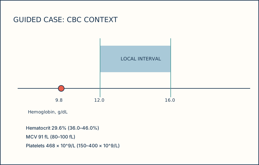
| Test | Result | Local interval |
|---|---|---|
| Hemoglobin | 9.8 g/dL | 12.0-16.0 g/dL |
| Hematocrit | 29.6% | 36.0-46.0% |
| MCV | 91 fL | 80-100 fL |
| Platelets | 468 × 10^9/L | 150-400 × 10^9/L |
Knowledge checks
Reading and checks are open. Sign in only to save.
Knowledge check 1
Section status
Finish this section
Reading and checks are open. Sign in only to save.
The module finishes after every required section is marked done and every check in those sections is correct.