Required section · Section 5 of 6
Classify evidence and choose the bounded action
A normal average MCV does not exclude a mixed red-cell population. Mixed iron and B12 or folate deficiencies can normalize MCV while increasing RDW. The histogram below illustrates a reference-range average MCV with separate small and large cell populations.
For the guided case, rank the macro-ovalocytic smear pattern, low reticulocytes, and functional-marker pattern above unsupported disease-name guessing. Pancytopenia requires qualified morphology review and escalation through the local morphology-review and critical-result escalation procedure. Dysplasia or blasts also require that escalation pathway.
Illustrative drawing — this picture was drawn rather than captured.
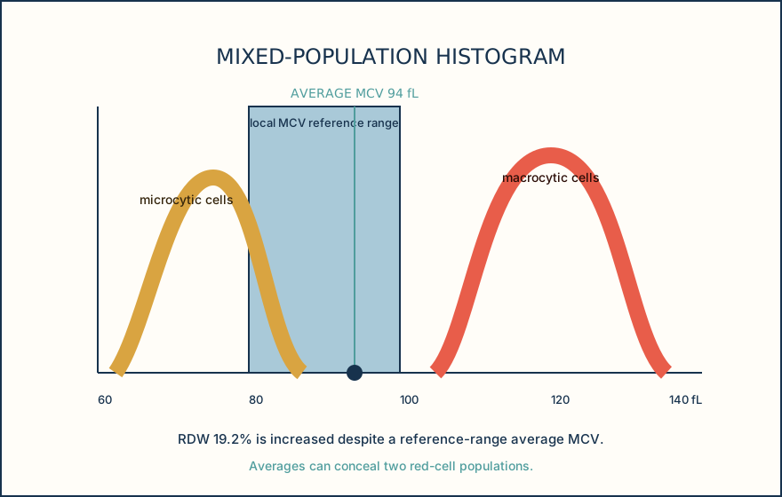
| Population | Approximate MCV | Note |
|---|---|---|
| Microcytic peak | 75 fL | Smaller red-cell subpopulation |
| Macrocytic peak | 120 fL | Larger red-cell subpopulation |
| Average MCV | 94 fL | Within the 80-100 fL reference band |
| RDW | 19.2% | Increased despite a reference-range average MCV |
Ordering exercise
Place the review actions in the sequence that protects the interpretation from a misleading MCV.
1. Correlate targeted markers
Interpret nutrient markers and chemistry with assay and renal context.
2. Review qualified morphology
Classify morphology and identify escalation findings.
3. Assess reticulocyte response
Determine whether regeneration may increase MCV.
4. Confirm CBC and specimen
Review specimen condition, analyzer flags, count, and hematocrit.
Knowledge checks
Reading and checks are open. Sign in only to save.
Knowledge check 1
Knowledge check 2
Section status
Finish this section
Reading and checks are open. Sign in only to save.
The module finishes after every required section is marked done and every check in those sections is correct.