Required section · Section 3 of 6
Focused instruction: pair methods and name limits
Cation-exchange HPLC separates fractions by retention time and can quantify HbA2 and HbF. Capillary electrophoresis separates by electrophoretic mobility and displays zones, giving a complementary principle. Retention-time and zone identifications remain method-specific. Review the analyzer, reagent application, calibrator, controls, software, and reportable fractions on the local method card before releasing an interpretive comment.
An S-position band on alkaline electrophoresis is not uniquely HbS because HbD, HbG, and Hb Lepore can co-migrate there. Acid electrophoresis can separate HbS from HbD and HbG in that setting. A major fraction in an S zone on capillary electrophoresis supports a sickling hemoglobin but does not genotype it. Molecular HBB testing or family studies can resolve remaining ambiguity under the local pathway.
A sickling-solubility screen observes turbidity when reduced sickling hemoglobin becomes insoluble in concentrated phosphate buffer. It is qualitative and does not distinguish sickle cell disease from trait. The SICKLEDEX IFU, document 350430-27 revision 2024-05, states a kit-specific 15% HbS detection limit at total hemoglobin 12 g/dL and cautions that high HbF below age six months and recent transfusion can affect results. Use solubility as a limited screen, never as the genotype result.
Illustrative drawing — this picture was drawn rather than captured.
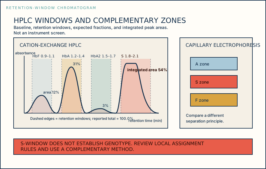
| Window | Retention range (min) | Reported area |
|---|---|---|
| HbF | 0.9–1.1 | 12.0% |
| HbA | 1.2–1.4 | 31.0% |
| HbA2 | 1.5–1.7 | 3.0% |
| S | 1.8–2.1 | 54.0% (integrated) |
Knowledge checks
Reading and checks are open. Sign in only to save.
Knowledge check 1
Knowledge check 2
Section status
Finish this section
Reading and checks are open. Sign in only to save.
The module finishes after every required section is marked done and every check in those sections is correct.