Required section · Section 3 of 6
High-concern clues and look-alikes
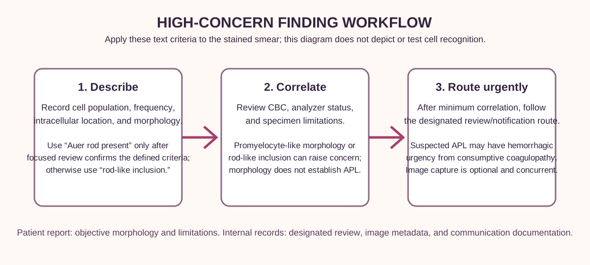
Report “Auer rod present” only after focused review identifies a sharply defined red to reddish-purple rod or needle-like azurophilic cytoplasmic inclusion in an immature myeloid-appearing cell. Use “rod-like cytoplasmic inclusion” when that morphology, intracellular location, cell context, or focused review is not sufficiently established. Either term is an objective morphology statement, not an acute myeloid leukemia or subtype diagnosis. Record the cell population and frequency; do not reduce the finding to a routine plus grade.
Hypogranularity or agranularity and hyposegmented pseudo-Pelger-Huët nuclei are dysgranulopoietic features. Compare a pseudo-Pelger-like neutrophil with a normally segmented neutrophil: the former has a bilobed or unilobed, often coarse-contoured nucleus, whereas the latter has multiple connected lobes. This descriptive morphology is not itself a myelodysplastic-neoplasm diagnosis and must be distinguished from inherited Pelger-Huët morphology using the clinical history and the laboratory pathway.
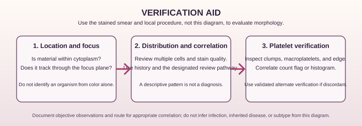
Morula-like material is a compact basophilic granular cluster within white-cell cytoplasm, not debris overlying or outside the cell. During the first week of illness, microscopy may show ehrlichial or anaplasmal morulae, but a smear lacks sensitivity for standalone diagnosis and cannot conclusively distinguish Ehrlichia from Anaplasma. Check intracellular location through the cell plane, focus behavior, distribution, stain quality, cell type, and CBC context before reporting an organism-like finding.
MYH9-related disease can show congenital thrombocytopenia, giant platelets, and neutrophil cytoplasmic MYH9 aggregates that can resemble Döhle bodies. Compare a reactive Döhle body with an MYH9-like aggregate across cells and platelet morphology: neither establishes MYH9-related disease from the smear. First verify the platelet result: assess the smear for clumping and macroplatelets, including the feathered edge; correlate the platelet histogram or flag; and use the laboratory's validated alternate count or estimate when large platelets may cause an electronic undercount. Document the observed pattern and route it for correlation rather than naming MYH9-related disease from the smear.
Illustrative drawing — this picture was drawn rather than captured.
Illustrative drawing — this picture was drawn rather than captured.
Knowledge checks
Reading and checks are open. Sign in only to save.
Knowledge check 1
Knowledge check 2
Knowledge check 3
Section status
Finish this section
Reading and checks are open. Sign in only to save.
The module finishes after every required section is marked done and every check in those sections is correct.