Required section · Section 3 of 6
Focused instruction: preserve evidence and use the right method
For case 1, preserve the remaining EDTA aliquot immediately, in parallel with running the CBC and smear rather than waiting until after they result, since a flow specimen's stability window begins at collection, not at the CBC report. Contact the receiving flow laboratory before assuming EDTA whole blood is a usable specimen for their validated method: some panels require a different anticoagulant, a fresh marrow specimen, or a narrower collection-to-receipt window than EDTA peripheral blood can meet.
Multiparameter flow cytometry may identify an abnormal precursor population, but trusting that finding requires four things in sequence: specimen suitability (correct anticoagulant, no clotting, adequate cellularity under the local method), viability at or above the local validated threshold, acquisition of at least the local target-event count in the region of interest, and a gating strategy that separates the population of interest from debris, doublets, and mature cells. When a limited peripheral-blood specimen fails suitability, viability, or target-event criteria, the required action is to report the specimen as unacceptable or limited for the requested testing, document the reason, and pursue recollection or a marrow specimen per local policy — never to report a percentage computed from an inadequate sample. Even a specimen that passes every gate still needs the full hematopathology workup: panel, fluorochromes, analyzer, software, and report language are local validated-method details.
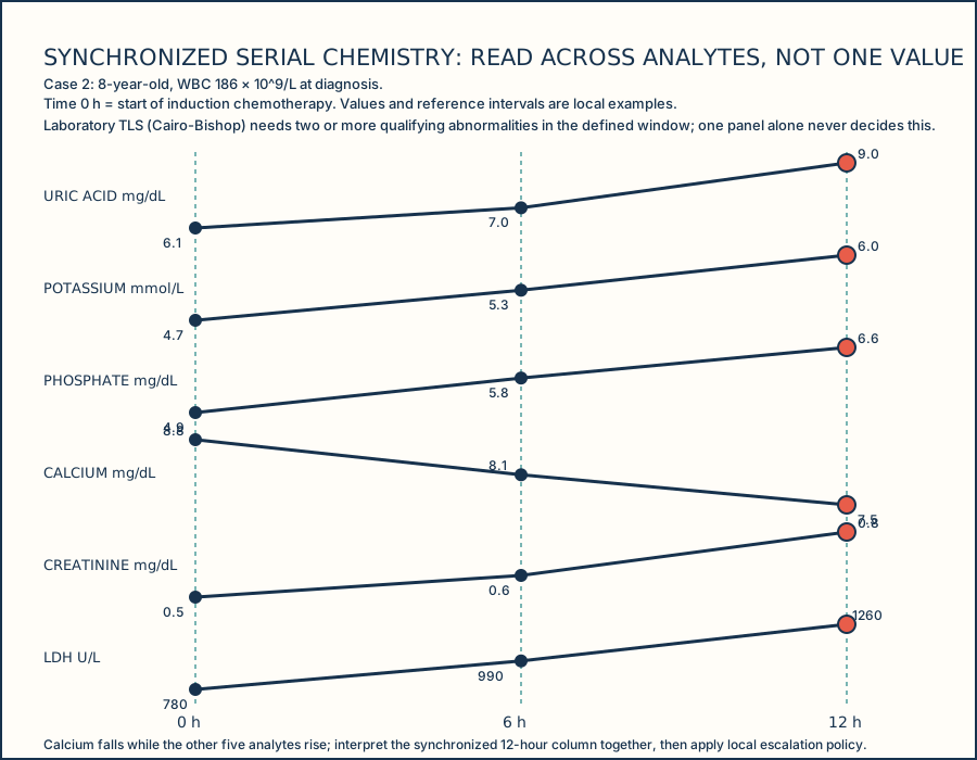
For case 2, an 8-year-old with newly diagnosed B-lymphoblastic leukemia presents with WBC 186 × 10^9/L (hyperleukocytosis) at diagnosis. Time 0 h marks the start of induction chemotherapy; serial chemistry is drawn at 0, 6, and 12 hours. Hyperleukocytosis and tumor-lysis chemistry require urgent attention under the approved local policy. Uric acid, potassium, phosphate, calcium, creatinine, and lactate dehydrogenase (LDH) contribute laboratory evidence, each read against a local reference interval.
Cairo-Bishop laboratory tumor lysis syndrome (TLS) criteria require two or more qualifying metabolic abnormalities (elevated uric acid, potassium, or phosphate, or decreased calcium) within the defined treatment-timing window, so one isolated result cannot settle the assessment. Clinical TLS adds a clinical consequence to that laboratory picture — a rising creatinine, a cardiac arrhythmia, or a seizure. Leukostasis is a separate hyperviscosity syndrome driven by a very high circulating blast count, producing respiratory or neurologic symptoms; it is mechanistically distinct from the metabolic derangements of TLS and can occur with or without laboratory or clinical TLS. Pretreatment WBC, LDH, and uric acid function as risk markers that inform TLS risk stratification — they raise or lower concern — but none of them is a diagnostic threshold by itself.
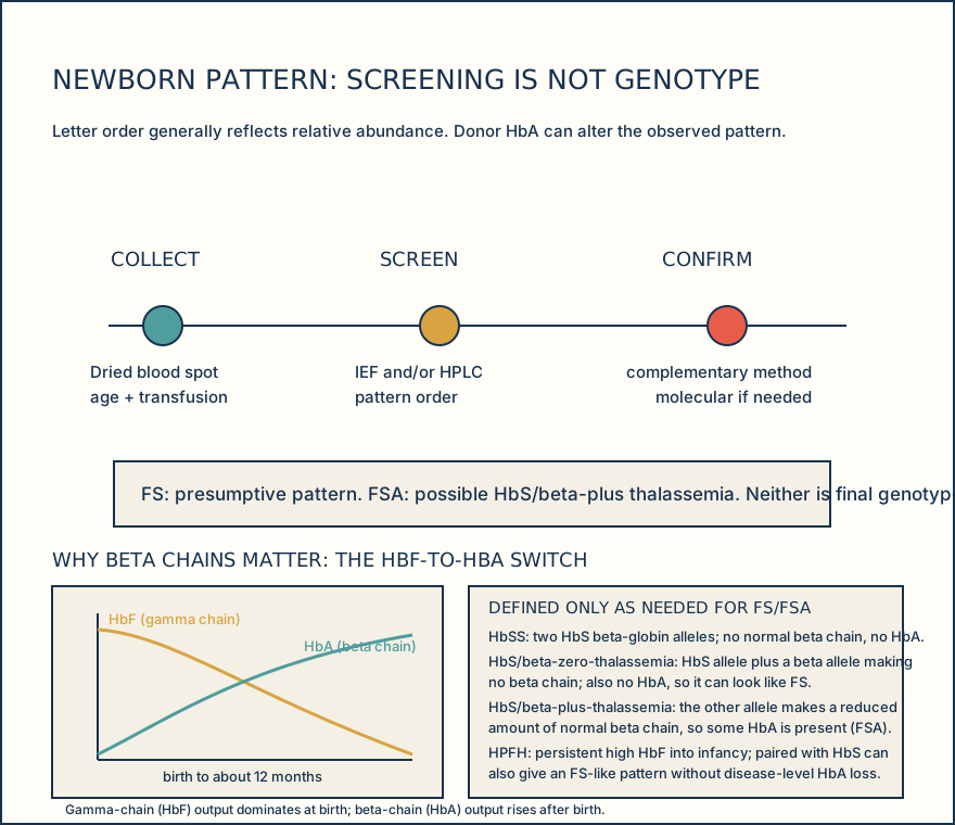
For newborn case 3, record collection age and transfusion history before interpreting FS or FSA. Both IEF and HPLC share a limitation that must be understood before checking a screen result against it: HPLC separates hemoglobins by chromatographic retention time, and two different variants can co-elute at the same or overlapping retention time, so retention time alone cannot identify every variant. IEF separates hemoglobins by isoelectric point, and two different variants can co-migrate to the same band position, so band position alone is not sufficient either. This is exactly why a complementary method, and molecular testing when needed, resolves what a single screening method cannot. FS is presumptive and can reflect HbSS, HbS/beta-zero thalassemia, or less commonly HbS with hereditary persistence of fetal hemoglobin. FSA is consistent with possible HbS/beta-plus thalassemia, not a final genotype.
Case 4 is a newborn screened on day 3 of life. The infant received a simple (non-exchange) transfusion of 15 mL/kg leukoreduced packed red cells on day 1, two days before the dried-blood-spot collection, for anemia of prematurity. The screen shows an FA pattern with a lower HbF fraction than expected for a 3-day-old infant, consistent with donor HbA diluting the infant's true baseline. Because the affected specimen was collected within the transfusion-affected interval, the jurisdictional algorithm calls for a repeat dried blood spot once donor red cells have cleared; CDC/APHL guidance describes repeating hemoglobinopathy screening around four months post-transfusion when a result is transfusion-affected, rather than accepting the transfusion-influenced pattern as the infant's baseline. CAP requires the collection manual to define preservation, transport, and storage conditions, and labeling and suboptimal-specimen handling criteria are defined locally. Preserve the specimen and record transfusion context before assigning meaning to a fraction pattern.
Illustrative drawing — this picture was drawn rather than captured.
Illustrative drawing — this picture was drawn rather than captured.
| Item | Value |
|---|---|
| Age | 8 years |
| Presenting WBC | 186 × 10^9/L (hyperleukocytosis) |
| Working diagnosis | Newly diagnosed B-lymphoblastic leukemia |
| Treatment anchor | Time 0 h = start of induction chemotherapy |
| Time | Uric acid | Potassium | Phosphate | Calcium | Creatinine | LDH |
|---|---|---|---|---|---|---|
| 0 h | 6.1 mg/dL | 4.7 mmol/L | 4.9 mg/dL | 8.8 mg/dL | 0.5 mg/dL | 780 U/L |
| 6 h | 7.0 mg/dL | 5.3 mmol/L | 5.8 mg/dL | 8.1 mg/dL | 0.6 mg/dL | 990 U/L |
| 12 h | 9.0 mg/dL | 6.0 mmol/L | 6.6 mg/dL | 7.5 mg/dL | 0.8 mg/dL | 1260 U/L |
| Local reference interval (example) | 2.5-5.5 mg/dL | 3.5-5.0 mmol/L | 3.0-4.5 mg/dL | 8.5-10.5 mg/dL | 0.3-0.7 mg/dL | 120-300 U/L |
Knowledge checks
Reading and checks are open. Sign in only to save.
Knowledge check 1
Knowledge check 2
Knowledge check 3
Knowledge check 4
Section status
Finish this section
Reading and checks are open. Sign in only to save.
The module finishes after every required section is marked done and every check in those sections is correct.