Required section · Section 3 of 6
Counting cells and reading the chemistry boundary
The total nucleated cell count and differential can be performed manually or with a validated automated method. Clinical and Laboratory Standards Institute (CLSI) document H56 covers collection, transport, processing, manual and automated enumeration, and reporting of cellular components in pleural and peritoneal fluid, and it expects the laboratory to use its own validated method rather than assume manual and automated counts are interchangeable. For manual counts specifically, each sample is counted in duplicate with defined limits of agreement between the two counts. Agreement between the two counts is what confirms the specimen was mixed well, loaded into the chamber without a bubble or a starved edge, and distributed evenly across the ruled area; a single chamber fill cannot rule any of those failures out on its own, because one fill can reflect a local clump, an air pocket, or uneven settling just as easily as it reflects the true cell concentration. When the two counts disagree beyond the defined limit, that disagreement is itself evidence of a mixing, loading, distribution, or counting error somewhere upstream, so the laboratory recounts rather than averaging the two values or reporting whichever one looks more plausible.
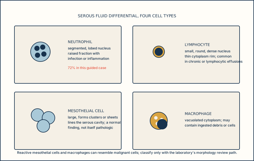
The differential reports the percentage distribution of identified cell types: neutrophils, lymphocytes, mesothelial cells, macrophages, and others. Mesothelial cells and macrophages are both larger than lymphocytes and can look alike at low power, but they separate on nucleus, border, size, and cytoplasm: a mesothelial cell has a round to oval, centrally placed nucleus with a smooth border and dense, non-vacuolated cytoplasm, often arranged in flat sheets with scalloped or window-like borders where adjacent cells crowd together; a macrophage has a more variable, often eccentric or kidney-bean-shaped nucleus and abundant foamy, vacuolated cytoplasm that may contain ingested debris. A malignant look-alike is called on the same features rather than a generic label: an irregular or thickened nuclear border, coarse or unevenly clumped chromatin, a nucleus disproportionately large for its cytoplasm, multiple or markedly enlarged nucleoli, or clusters of cells with molded, overlapping borders instead of the flat, evenly spaced sheets typical of reactive mesothelial cells. Any of those findings routes the slide to cytology or pathology rather than being called benign on the differential alone. The absolute neutrophil count is then calculated as total nucleated cells multiplied by the neutrophil fraction. The method used to prepare the differential slide matters: stained cytocentrifuge preparations give better cell yield and morphologic detail than unstained hemocytometer preparations, which are considered suboptimal outside limited circumstances such as distinguishing polymorphonuclear from mononuclear cells. A laboratory that uses an alternate method must show it is equivalent before relying on it for sub-classification or malignant-cell detection.
Pleural fluid classification most often uses Light's criteria: a fluid is called an exudate if any one of three conditions is met, fluid-to-serum protein ratio greater than 0.5, fluid-to-serum LDH ratio greater than 0.6, or fluid LDH greater than two-thirds of the serum LDH upper reference limit. These criteria classify pleural fluid only. They are not a peritoneal fluid rule, and applying them to ascites would misclassify the specimen. For serum and fluid protein and LDH to make a valid pair, both should be drawn close to the thoracentesis so the ratio reflects one clinical moment rather than two different physiologic states.
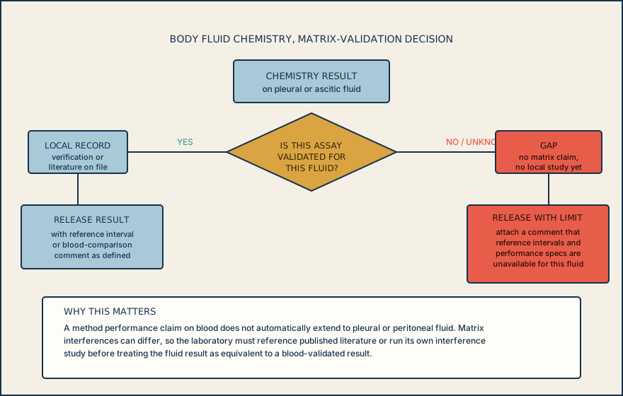
Not every chemistry assay on the menu is validated for a body fluid matrix, and two different gaps get confused at the bench. The first is a missing reference interval: the method has already been verified to perform accurately on pleural or peritoneal fluid, with matrix interference reasonably excluded either by citing published literature in the procedure or by running the laboratory's own interference study, but no fluid-specific reference interval has been established. That result can be released, with a comment that a fluid-specific reference interval is unavailable so the number is read against trend or the clinical picture rather than a numeric range. The second gap is missing matrix validation itself: the laboratory has not established or verified that the method's performance specifications apply to this fluid at all. Under 42 CFR 493.1253, a laboratory must establish or verify performance specifications for a test system before reporting patient results with it, and that requirement covers the specimen type being tested, not only blood. A comment does not substitute for that verification; an unvalidated matrix result is not made releasable by attaching a disclaimer. Until the interference study or equivalent verification is complete, the result is not reported from this method for this fluid, and the specimen is referred to a method or a laboratory that has already completed that validation.
Know whether today's chemistry result is missing a fluid-specific reference interval or missing matrix validation altogether. The first can be released with a comment describing the gap; the second cannot be released at all until the laboratory completes that verification.
Illustrative drawing — this picture was drawn rather than captured.
Illustrative drawing — this picture was drawn rather than captured.
| Aliquot | Destination test | Common failure | Consequence if missed |
|---|---|---|---|
| EDTA, mixed | Cell count and differential | Clot or poor mixing | Falsely low or inconsistent count |
| Sterile, no anticoagulant | Microbiology and cytology | Contamination or insufficient volume | Lost culture or cytology yield |
| Plain chemistry | Protein, LDH, glucose, triglyceride, amylase | No paired serum drawn | Cannot calculate Light's criteria ratios |
| Heparinized blood-gas syringe | pH | Air exposure or delayed run | pH artifact can shift the risk band |
Knowledge checks
Reading and checks are open. Sign in only to save.
Knowledge check 1
Knowledge check 2
Section status
Finish this section
Reading and checks are open. Sign in only to save.
The module finishes after every required section is marked done and every check in those sections is correct.