Required section · Section 1 of 6
The specimen on the bench
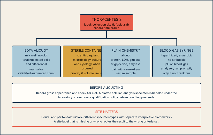
A thoracentesis specimen arrives at 10:10 labeled left pleural fluid, collected at 10:02. It is opaque yellow with no visible clot. The requisition asks for a cell count and differential, protein, LDH, glucose, and pH, plus culture. Before any of those results mean anything, the fluid has to be split into the right containers, and the site label on the tube has to match the site the physician wrote on the order.
Pleural fluid and peritoneal fluid look alike on the bench, but they come from different cavities and are governed by different interpretive frameworks. A tube labeled only fluid, with no site, cannot be safely classified once it leaves the collection area. That single label decision determines which criteria apply to every downstream calculation.
One guided pleural case is worked end to end: routing the specimen, reading its gross appearance, counting and differentiating the cells, and calculating the ratios that classify it as an exudate or transudate. It also marks where a pleural rule stops applying, where peritoneal fluid needs a different framework, and where a chemistry result depends on a validation study the reader cannot see from the bench.
Read the order and the tube label together before you aliquot anything; a fluid without a confirmed collection site is not yet ready for a body-fluid protocol.
Illustrative drawing — this picture was drawn rather than captured.
Knowledge checks
Reading and checks are open. Sign in only to save.
Knowledge check 1
Section status
Finish this section
Reading and checks are open. Sign in only to save.
The module finishes after every required section is marked done and every check in those sections is correct.