In an inhibition assay, no clumping is the positive result, and a sandwich immunoassay can read falsely low when the analyte is very high. Serologic procedures convert an antigen-antibody reaction into a visible, enzymatic, fluorescent, or electronic signal. Each format determines how a result is interpreted, which controls show that the run is valid, and which interferences can affect the result. Laboratory Operations covers general quality assessment and the sensitivity, specificity, and predictive-value calculations used in interpretation. Chemistry covers measurement of analyte-specific immunoassays.
Precipitation and agglutination
The lattice principles behind precipitin lines, reaction zones, and titer interpretation are covered in Antigen-Antibody Interactions and Complement.
Precipitation forms a visible lattice from soluble antigen and antibody. Ouchterlony double immunodiffusion places antigen and antisera in wells cut into an agarose gel and lets each diffuse toward the other. Precipitin lines form where concentrations reach equivalence, and comparing control and unknown wells gives three patterns: fused, continuous lines show identity, crossing lines show nonidentity, and a fused line with a spur shows partial identity, where one antigen shares an epitope with a second and has an additional one. Anti-Sm and anti-U1 RNP can produce partial identity because their targets occur in related small nuclear ribonucleoprotein complexes but are not identical. Immunodiffusion remains a reference method for resolving which extractable nuclear antigen an antibody recognizes, with high specificity and low throughput.1,2
Flocculation is precipitation confined to a narrow antigen-concentration window. The VDRL and RPR flocculation assays apply it to lipoidal antigens as described in Syphilis, Lyme, and Tuberculosis Serology.1
Agglutination applies the same cross-linking to particulate antigen, including whole cells and inert particles coated in advance with antigen or antibody.
Scroll sideways to see all columns
| Reaction type | Setup | Positive result means |
|---|---|---|
| Direct agglutination | Patient serum reacts with antigen naturally present on the particle | Antibody to that native antigen is present |
| Passive (indirect) agglutination | Patient serum is mixed with inert particles coated with antigen | Antibody to the attached antigen is present |
| Reverse passive agglutination | Patient sample is mixed with particles coated with reagent antibody | The corresponding antigen is present in the sample |
| Agglutination inhibition | Soluble hapten in the sample competes with hapten-coated particles for limited reagent antibody | No agglutination means antigen or hapten is present |
| Hemagglutination inhibition | Patient antibody neutralizes a virus that would otherwise bridge red cells into a lattice | No agglutination means antibody is present |
In the inhibition formats, no agglutination is the positive result. Cryptococcal antigen testing by latex agglutination uses the reverse-passive format. Lateral-flow cryptococcal antigen assays are generally preferred where available because they are rapid and simple.1
On an RPR card, antilipoidal antibody in the specimen binds the cardiolipin antigen into a lattice that traps the charcoal particles. The visible clumps are called floccules.15 A reactive specimen shows black floccules against the white card. A nonreactive specimen shows a uniform light-gray color. Reactive, minimally reactive, and nonreactive controls are tested in each run, and patient results are reported only when every control gives its expected pattern.16
Hemagglutination is read in a microtiter well. Agglutinated red cells do not settle and coat the well as a diffuse film. Red cells that do not agglutinate settle into a compact button. In hemagglutination inhibition, a button in a patient well means antibody inhibited viral hemagglutination at that dilution. That reading is valid only when the cell control without virus settles into a button and the positive-control antiserum inhibits as expected.17
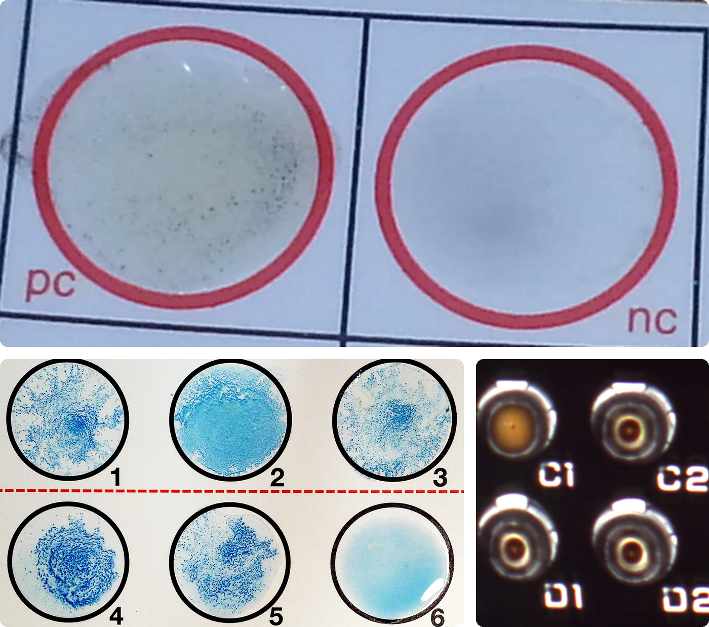
Agglutination and precipitation assays are fast, portable, inexpensive, and instrument-free, which suits them to pathogens that are dangerous or slow to culture. Antigen concentration, incubation time and temperature, diluent, and reading method all shift the equivalence point. Run them exactly as validated, use reagents within their validated life, and verify each new reagent lot, because kit sensitivity and specificity differ across manufacturers.1,16 For an unexpected reactive result, verify specimen identity and assay validity and complete the assay's supplemental algorithm. When the combined results or clinical context remain discordant, investigate cross-reactivity, in which an antibody binds a structurally similar epitope, or another interference. Monoclonal reagents directed at a single epitope reduce the risk of cross-reactivity. A negative result does not exclude analyte below the detection threshold, so repeat a negative early-window serologic test on the schedule its algorithm defines.1
Labeled immunoassays
Labeled immunoassay formats differ in two design features: how bound and free label are separated, and whether patient analyte competes with the reagent or is captured between two antibodies.1
Separation. Heterogeneous assays physically separate bound from free label with a wash, magnetic capture, or centrifugation before reading. Homogeneous assays are designed so that bound and free label give different signals in the same tube. This design sacrifices some analytical sensitivity for speed and full automation.1
Binding format. Competitive assays give labeled and patient analyte a fixed, limited number of antibody sites, so more patient analyte leaves less bound label: when bound label is measured, its signal decreases as patient analyte increases. Homogeneous competitive formats such as the enzyme multiplied immunoassay technique (EMIT) and the cloned enzyme donor immunoassay (CEDIA) measure an enzyme signal that rises with analyte. The design suits small haptens that cannot bind two antibodies at once. Sandwich (capture) assays use capture antibody in excess across the validated measuring interval, allowing a large fraction of patient analyte to bind before a labeled second antibody against a different epitope completes the sandwich. The signal rises directly with analyte within that interval, and the format requires a large enough analyte to present two distinct epitopes.1
In an idealized competition model, labeled and patient antigen have equal affinity and access to the same binding sites. If binding does not substantially deplete either free pool, the labeled share of bound antigen follows their relative abundance. With 90 units of labeled antigen, the model predicts:
Scroll sideways to see all columns
| Patient antigen | Labeled-to-patient ratio | Total antigen | Bound-label fraction |
|---|---|---|---|
| 30 units | 3 to 1 | 120 | 90 ÷ 120 = 75.0% |
| 90 units | 1 to 1 | 180 | 90 ÷ 180 = 50.0% |
| 270 units | 1 to 3 | 360 | 90 ÷ 360 = 25.0% |
These percentages describe the labeled share of the bound antigen. They do not show what fraction of all labeled antigen was captured. Actual assays use calibrators at known concentrations and an assay-specific curve fit to account for binding and signal behavior.1
Historical and current formats
Scroll sideways to see all columns
| Format | Label and principle | Reading | Laboratory note |
|---|---|---|---|
| RIA (radioimmunoassay) | Radioisotopes (3H, 14C, 125I, 131I); competitive | Counts per minute | First practical peptide-hormone immunoassay (Yalow and Berson, insulin, published 1960; Nobel Prize 1977); now largely historical because of radiation safety, waste handling, and short isotope shelf life |
| EIA/ELISA (enzyme immunoassay, enzyme-linked immunosorbent assay) | Enzyme (horseradish peroxidase, alkaline phosphatase, beta-D-galactosidase) converts many substrate molecules, amplifying signal | Color or absorbance | Indirect ELISA detects patient antibody on adsorbed antigen; sandwich ELISA detects antigen between capture and labeled antibody; used for antibodies to hard-to-culture pathogens and autoantibodies |
| CLIA (chemiluminescent immunoassay) | Chemiluminescent oxidation (acridinium ester, ruthenium, luminol) | Photons | High analytical sensitivity and broad dynamic range; performance and dilution requirements remain assay specific |
| CMIA (chemiluminescent microparticle immunoassay) | Acridinium chemistry on magnetic microparticles | Photons from particles | Heterogeneous despite solution-like handling |
| ECLIA (electrochemiluminescence immunoassay) | Ruthenium-labeled antibody oxidized electrochemically at an electrode | Electrochemiluminescence | High sensitivity on automated platforms |
| EMIT, CEDIA | Homogeneous enzyme assays for haptens | Enzyme activity restored as patient hapten displaces the labeled fragment | EMIT: antibody binding sterically blocks the enzyme active site; CEDIA: antibody-sequestered beta-galactosidase fragments recombine when displaced; both raise signal with analyte, so they are competitive homogeneous assays |
| FPIA (fluorescence polarization immunoassay) | Fluorescent hapten; bound hapten rotates too slowly to depolarize light | Polarization | Polarization falls as patient analyte rises; displaced by chemiluminescence on most platforms |
| Immunochromatographic | Colloidal gold or latex conjugate wicks along a nitrocellulose strip | Visible line | Noncompetitive tests show a line for a positive result (urine hCG, rapid strep, troponin); in a valid competitive drug screen a test line indicates a negative result at the assay's cutoff, so drug may still be present below that cutoff, and an absent test line with a valid control line indicates a presumptive positive result; the control line must appear for any result to be valid |
An indirect ELISA coats the well with antigen, adds patient serum, washes, adds enzyme-labeled anti-human immunoglobulin, washes again, and then adds substrate before the color is read. A sandwich ELISA starts with capture antibody, adds the patient sample, washes, adds an enzyme-labeled detection antibody, washes again, and then adds substrate. The historical Western blot separates antigens by electrophoresis, transfers them to a membrane, adds patient serum, washes, adds labeled anti-human IgG, washes again, and develops the band pattern with substrate. Each wash removes unbound reagent and limits background.1
Biotin-streptavidin amplification, with biotin on the detection antibody and enzyme-conjugated streptavidin recruiting multiple biotin sites, increases sandwich-assay sensitivity.1
Interferences
Scroll sideways to see all columns
| Mechanism | Effect | Typical fix |
|---|---|---|
| High-dose hook (antigen excess) | Sandwich assays read falsely low | Dilute and retest; confirm that the dilution-corrected result increases |
| Rheumatoid factor (anti-IgG) | Bridges assay antibodies; false positive, mainly in sandwich formats | Investigate with a validated blocking procedure, dilution study, or an alternate assay; no blocker works for every antibody interference4 |
| Heterophile and human anti-mouse antibodies | Cross-link capture and detection antibody directly; usually false positive, occasionally false negative | Blocking reagent or an orthogonal method |
| Biotin at high dose | Competes with the biotinylated reagent for streptavidin; false increase in competitive assays, false decrease in sandwich assays | Ask about dose, timing, kidney function, and whether the assay uses streptavidin-biotin; delay collection per the manufacturer's washout guidance, which may extend to 72 hours or beyond with high doses and renal impairment |
| Cross-reactivity | A structurally similar molecule is measured as the analyte | Confirm unexpected positives with a chemically orthogonal method |
FDA's safety communication warns that high-dose biotin from supplements marketed for hair, skin, and nails has caused falsely low troponin results and other serious misinterpretations in streptavidin-biotin assays, and the agency asks patients to disclose biotin use before testing.3 Immunoassays are not standardized across manufacturers because kits target different epitopes, so reference intervals and absolute values differ by platform. Run serial specimens for one patient by one method.1
Immunofluorescence and multiplex bead assays
In direct immunofluorescence, the fluorochrome is conjugated to the antibody that recognizes the target. Antigen fixed to a slide, or present on cells, is incubated with the labeled antibody, washed, and read under ultraviolet illumination. Bright fluorescence on a dark field is positive. The format identifies microbial antigen directly in specimens, with Legionella pneumophila and Chlamydia trachomatis as classical applications, and, read by flow cytometry, it is the basis of leukemia and lymphoma immunophenotyping with CD-marker panels.1
Indirect immunofluorescence (IIF) adds an amplifying second layer: fixed antigen or cells capture patient antibody, then a fluorescent anti-human-immunoglobulin conjugate binds it. Several labeled secondary molecules can bind each captured primary antibody, so IIF amplifies signal beyond direct labeling, and one anti-human conjugate can be used with every antigen substrate in the laboratory. For antigen detection, IIF uses an unlabeled primary antibody first. For antibody detection, patient antibody acts as the primary reagent. It remains the reference method for HEp-2 ANA screening, Crithidia luciliae anti-dsDNA confirmation, the historical fluorescent treponemal antibody absorption (FTA-ABS) test, varicella-zoster virus (VZV) FAMA testing, and Epstein-Barr virus (EBV)-specific serology. It is slow, requires a trained reader, and varies with buffer, conjugate, and microscope conditions among laboratories.1
Multiplex bead immunoassays dye polystyrene beads with unique ratios of red and infrared fluorescent dyes, giving 100 to 500 distinct identities in commercial platforms, one antigen or one antibody per bead. Flow cytometry reads each bead's color identity and bound-antibody fluorescence simultaneously, so single-antigen HLA antibody testing, autoantibody panels, donor-specific antibody screening, and cytokine profiling run from one small sample volume.1
Autoantibody serology
Antinuclear antibodies
Antinuclear antibodies target nuclear components, including dsDNA, histones, nucleosomes, and centromere proteins. Extractable nuclear antigen panels include Sm, RNP, SS-A/Ro, SS-B/La, and other named specificities. Some panels also include anti-Jo-1, whose target is cytoplasmic. ANA is present in most patients with systemic lupus erythematosus and also occurs in other autoimmune diseases, chronic infections, some cancers, pregnancy, and healthy people. Prevalence in healthy populations rises with age and with lower screening dilutions, so a percentage applies only to the stated method, cutoff, and population. A positive ANA is therefore a sensitive screen with limited specificity.1
Scroll sideways to see all columns
| Autoantibody | Antigen | IIF pattern | Chief association |
|---|---|---|---|
| Anti-dsDNA | Double-stranded DNA | Homogeneous | Supports SLE; assay-dependent specificity and activity correlation |
| Anti-histone | Histones H1 to H4 | Homogeneous | Drug-induced lupus |
| Anti-nucleosome | DNA-histone complex | Homogeneous | SLE association; sensitivity and activity correlation depend on the assay and population |
| Anti-Sm | Uridine-rich RNA-bound protein | Coarse speckled | SLE-specific; limited sensitivity |
| Anti-RNP | U1-snRNP | Coarse speckled | High levels support mixed connective tissue disease in the appropriate clinical pattern |
| Anti-SS-A/Ro and anti-SS-B/La | RNA-bound proteins | Finely speckled | Sjögren disease and SLE; maternal antibodies are associated with neonatal lupus |
| Anti-Scl-70 | DNA topoisomerase I | Topo I-like (AC-29) | Systemic sclerosis |
| Anti-Jo-1 | Histidyl-tRNA synthetase | Cytoplasmic fine speckling may occur; HEp-2 screening can miss it | Antisynthetase syndrome, including inflammatory myopathy and interstitial lung disease5,6 |
| Anti-centromere | CENP-A/B/C | Discrete nuclear dots with a characteristic metaphase pattern | Limited cutaneous systemic sclerosis, CREST |
| Anti-DFS70 | Transcription co-activator p75 | Dense fine speckled (AC-2) | A confirmed specificity is reassuring only with low clinical probability and appropriate assessment for other antibodies5 |
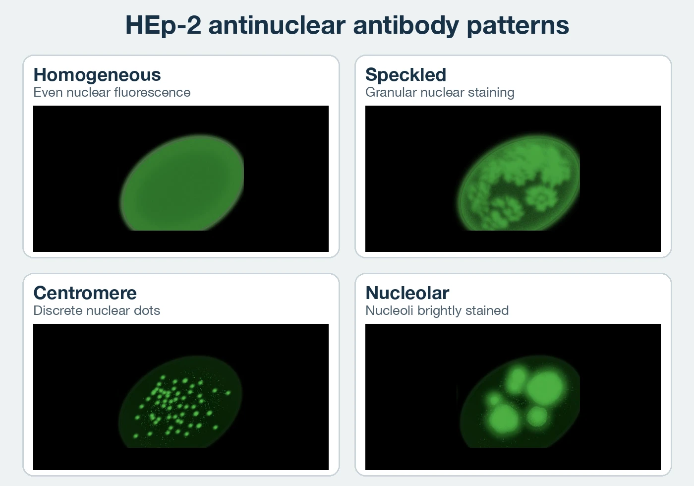
IIF on HEp-2 cells remains the reference ANA screening method: patient serum is incubated on fixed HEp-2 cells, chosen for large nuclei with high antigen density, developed with fluorescein-labeled anti-human immunoglobulin, and read at 400×, usually at screening dilutions of 1:80 to 1:160. The report states the endpoint titer and pattern, and the International Consensus on ANA Patterns (ICAP) standardizes the nomenclature. The current ICAP reference recognizes 31 anti-cellular patterns across nuclear, cytoplasmic, and mitotic categories, coded AC-1 through AC-31 plus AC-XX for unassigned patterns.1,8 A negative ANA result lowers the probability of SLE, but follow-up still depends on assay sensitivity and the clinical presentation.1
A nuclear dense fine speckled pattern (AC-2) requires careful recognition and antigen-specific confirmation of anti-DFS70. Its association with a lower likelihood of systemic autoimmune rheumatic disease applies chiefly when clinical suspicion is low. Anti-Ro52 and anti-Jo-1 may produce no positive HEp-2 screening result. Strong clinical suspicion therefore warrants the relevant antigen-specific testing even after anti-DFS70 is confirmed. The laboratory reports the pattern and confirmed specificity separately and completes clinically directed follow-up. The pattern and the ANA titer cannot substitute for an antigen-specific assay that was not performed.5
At the reporting level, a homogeneous pattern gives even diffuse fluorescence across each interphase nucleus. A speckled pattern gives granular nuclear staining. A centromere pattern gives about 40 to 80 discrete interphase dots and a characteristic metaphase plate pattern. A nucleolar pattern concentrates bright homogeneous or speckled staining in the nucleoli, with weak or absent nucleoplasmic staining.8
Solid-phase assays (ELISA, chemiluminescent, multiplex bead) anchor a defined nuclear antigen panel to a plate, membrane, or bead and read bound antibody with labeled anti-human immunoglobulin. They are faster and more objective than IIF but less sensitive as a screen, so professional guidance retains IIF as the reference screening method. Crithidia luciliae immunofluorescence confirms anti-dsDNA specifically: the kinetoplast of this trypanosome is an organelle of nearly pure dsDNA, so bright kinetoplast staining gives a specific signal free of the ssDNA reactivity that contaminates other substrates.1
Thyroid autoantibodies
Thyroid autoantibodies help identify the cause of thyroid dysfunction. Anti-thyroid peroxidase (anti-TPO) is associated with Hashimoto thyroiditis. Anti-thyroglobulin can provide additional evidence. TSH-receptor antibodies (TRAbs), particularly stimulating antibodies, support Graves disease. Antibody positivity does not establish the current biochemical state, which is assessed with thyroid-stimulating hormone (TSH) and thyroid hormone measurements. Detection rates depend on the assay and patient population.9
TRAb binding assays detect receptor-binding antibodies without separating stimulating from blocking activity. Functional bioassays measure the effect on receptor-bearing cells. A report identifies the assay and whether it measures receptor binding or stimulating activity. Do not infer the format from the test name alone.9
Rheumatoid factor and anti-CCP
Rheumatoid factor (RF) commonly represents IgM directed against the Fc region of IgG. It can occur in rheumatoid arthritis, other autoimmune diseases, infections, and healthy people. A negative result does not exclude rheumatoid arthritis, and a positive result requires the joint findings and clinical history for interpretation.10
Anti-cyclic citrullinated peptide (anti-CCP) antibody is generally more specific for rheumatoid arthritis than RF and can be present when RF is negative. Classification integrates RF and anti-CCP with joint involvement, symptom duration, and inflammatory markers. Erythrocyte sedimentation rate (ESR) and C-reactive protein (CRP) provide nonspecific evidence of inflammation. Antibody results alone do not establish current disease activity.10
Cytokine assays
Three assay families are used for clinical cytokine profiling for autoimmune disease, infection, allergy, and inflammatory states. Multiplexed ELISAs and microarrays measure several cytokines from one sample, each in its own well or array spot with paired controls. Microbead assays measure up to about 100 analytes per tube with colored, antigen- or antibody-coated beads read by flow cytometry, using the multiplex principle described above. Measured analytes include acute-phase reactants such as CRP, Th1 and Th2 discriminators such as IFN-gamma, IL-2, IL-4, IL-5, and IL-10, and colony-stimulating factors.1
ELISpot counts individual cytokine-secreting cells. The other two families measure total cytokine mass. Peripheral blood mononuclear cells isolated by gradient centrifugation are plated on capture-antibody-coated wells and stimulated with antigen. A sandwich-format detection step then leaves a countable spot of captured cytokine directly beneath each responding cell. T-SPOT.TB applies this principle to M. tuberculosis exposure, as described in Syphilis, Lyme, and Tuberculosis Serology.1
Test result interpretation
Confirmatory testing after screening
Screening and supplemental assays have different purposes, so the laboratory follows the validated algorithm for that analyte. Several infectious-disease algorithms use a different method or antigen set after a reactive screen: HIV screening reflexes to antibody differentiation, syphilis reflexes across the nontreponemal (lipoidal antigen) and treponemal divide, and Lyme reflexes from a first-tier immunoassay to an immunoblot or paired EIA. A solid-phase anti-dsDNA result may be followed by Crithidia luciliae immunofluorescence when the laboratory's algorithm calls for greater specificity.1
Complement interpretation
CH50 and AH50 pathway screens, their preanalytic requirements, and the follow-up of abnormal screens with component-specific antigenic and functional testing are covered in Antigen-Antibody Interactions and Complement. Loss of one C1 subcomponent can leave the other two antigenically present with no C1 function, and C2 antigen can be present without function, so antigenic measurement alone cannot exclude a functional defect.1
Immunodeficiency confirmatory testing
When screening or clinical findings suggest immunodeficiency, confirmatory tests narrow the defect to a compartment: humoral defects prompt B-cell counts by flow cytometry and in vitro B-cell proliferation testing; cell-mediated defects prompt T-cell subset counts, lymphocyte proliferation to mitogens and recall antigens, and adenosine deaminase and purine-nucleoside phosphorylase enzyme assays; phagocyte defects prompt adhesion-molecule analysis (CD11a/b/c, CD18), phagocytosis and killing assays, chemotaxis, and myeloperoxidase, G6PD, and NADPH-oxidase component assays; complement defects are resolved with the component assays described in the Antigen-Antibody Interactions and Complement lesson.11
Flow cytometry is central to that workup: fluorescent antibodies against CD markers on peripheral blood mononuclear cells after red-cell lysis identify the reduced CD3 population of partial DiGeorge syndrome, the profound T-cell deficiency of complete athymia, and markedly reduced B cells in classic BTK deficiency. Secondary immunodeficiency complicates interpretation. An absent B-cell result occurs after rituximab, an anti-CD20 monoclonal that depletes CD19-positive and CD20-positive B cells, so review the medication history before genetic testing is pursued. Interferon-gamma release assay (IGRA) platforms measure M. tuberculosis-specific responses and are not general cellular-immunity tests, and commercial ATP-release assays used in some transplant settings are not validated substitutes for standard inborn-error-of-immunity functional testing. Molecular testing is recommended for suspected SCID and many inborn errors of immunity. The laboratory reports urgent screening findings and identifies molecular confirmation as pending so the clinical immunology service can act under its protocol.11
General principles of serologic diagnosis
Serology detects the host response, so timing and interpretation are specific to the infection and assay. No single IgM timeline or paired-titer rule applies to all infections. For Lyme disease, early antibody testing can be negative and IgM results after more than 30 days of illness are disregarded. For EBV, viral capsid antigen (VCA) IgM with absent Epstein-Barr nuclear antigen (EBNA) antibody supports primary infection. Paired acute and convalescent sera usually add little. For hepatitis C, a reactive antibody result requires RNA testing to establish current infection.12-14
Each report identifies the test performed, its result, any incomplete supplemental testing, and the relevant timing limitation, so the reader knows the limits of the result. The infection-specific sequences are covered in Syphilis, Lyme, and Tuberculosis Serology, Viral Infectious Disease Serology, and HIV Testing and Monitoring.
Practice
References
- Abbas AK, Lichtman AH, Pillai S, Henrickson S. Cellular and Molecular Immunology. 11th ed. Elsevier; 2025. Elsevier. Back to text
- Benito-Garcia E, Schur PH, Lahita R; American College of Rheumatology Ad Hoc Committee on Immunologic Testing Guidelines. Guidelines for immunologic laboratory testing in the rheumatic diseases: anti-Sm and anti-RNP antibody tests. Arthritis Rheum. 2004;51(6):1030-1044. doi:10.1002/art.20836 Back to text
- US Food and Drug Administration. The FDA warns that biotin may interfere with lab tests: FDA safety communication. November 5, 2019. Accessed August 31, 2026. https://www.fda.gov/media/127915/download Back to text
- Ghazal K, Brabant S, Prie D, Piketty ML. Hormone immunoassay interference: a 2021 update. Ann Lab Med. 2022;42(1):3-23. doi:10.3343/alm.2022.42.1.3 Back to text
- International Consensus on ANA Patterns. AC-2: nuclear dense fine speckled. Updated August 2025. Accessed September 7, 2026. https://www.anapatterns.org/view_pattern.php?pattern=2 Back to text
- Castro C, Gourley M. Diagnostic testing and interpretation of tests for autoimmunity. J Allergy Clin Immunol. 2010;125(2 suppl 2):S238-S247. doi:10.1016/j.jaci.2009.09.041 Back to text
- ARUP Laboratories. Connective tissue diseases. ARUP Consult. Accessed September 7, 2026. Back to text
- Damoiseaux J. The International Consensus on ANA Patterns (ICAP): from conception to implementation. Clin Chem Lab Med. 2024;62(5):789-792. doi:10.1515/cclm-2023-1211 Back to text
- ARUP Laboratories. Autoimmune thyroiditis. ARUP Consult. Accessed September 7, 2026. Back to text
- ARUP Laboratories. Rheumatoid arthritis. ARUP Consult. Accessed September 7, 2026. Back to text
- Orange JS, Chinen J, Horner CC, et al. 2025 inborn errors of immunity practice parameter: guidance from the Joint Task Force on Practice Parameters, the American Academy of Allergy, Asthma & Immunology, the American College of Allergy, Asthma and Immunology, and the Clinical Immunology Society. Ann Allergy Asthma Immunol. 2026;136(4):426-493.e1. doi:10.1016/j.anai.2025.10.026 Back to text
- Centers for Disease Control and Prevention. Clinical testing and diagnosis for Lyme disease. Accessed September 7, 2026. https://www.cdc.gov/lyme/hcp/diagnosis-testing/index.html Back to text
- Centers for Disease Control and Prevention. Laboratory testing for Epstein-Barr virus. Accessed September 7, 2026. https://www.cdc.gov/epstein-barr/php/laboratories/index.html Back to text
- Centers for Disease Control and Prevention. Clinical screening and diagnosis for hepatitis C. Accessed September 7, 2026. https://www.cdc.gov/hepatitis-c/hcp/diagnosis-testing/index.html Back to text
- Papp JR, Park IU, Fakile Y, Pereira L, Pillay A, Bolan GA. CDC laboratory recommendations for syphilis testing, United States, 2024. MMWR Recomm Rep. 2024;73(1):1-32. doi:10.15585/mmwr.rr7301a1 Back to text
- Remel. RPR Card Test Instructions for Use. IFU 16302. Revised April 27, 2009. Accessed October 2, 2026. https://documents.thermofisher.com/TFS-Assets/MBD/Instructions/IFU16302.pdf Back to text
- Kaufmann L, Syedbasha M, Vogt D, et al. An optimized hemagglutination inhibition (HI) assay to quantify influenza-specific antibody titers. J Vis Exp. 2017;(130):55833. doi:10.3791/55833 Back to text