Required section · Section 3 of 6
What each test measures, and four look-alikes
PT and aPTT are plasma clot-initiation assays run on citrated platelet-poor plasma. PT measures the time to clot after tissue factor and calcium are added; aPTT measures the time to clot after a contact activator, phospholipid, and calcium are added. Both are one-stage clotting-time methods and, per CLSI H47, are sensitive to preexamination, reagent, and analytical variables. Neither test measures platelet contribution to clotting, full thrombin generation, or fibrinolysis, so a normal PT and aPTT do not rule out an abnormality in those other compartments.
Clauss fibrinogen adds excess thrombin to diluted plasma and converts the observed clot time to a concentration through a calibration curve; it is a functional assay, not a direct measurement of fibrinogen mass, so an abnormal fibrinogen molecule (dysfibrinogenemia) can distort the result in either direction.
D-dimer is generated when plasmin degrades cross-linked fibrin, so an increased result supports that fibrin was formed and then broken down. It is not specific for DIC. D-dimer can rise with thrombosis, infection, inflammation, surgery, trauma, pregnancy, malignancy, and liver disease, among other settings, so an isolated elevated D-dimer in an inflamed or postoperative patient carries limited discriminating value on its own; the trend and the company it keeps matter more than the single number.
Specimen integrity governs whether any of these results can be trusted. CLSI H21 sets the requirements for citrate tube fill, transport time and temperature, plasma preparation, and rejection criteria; hemolysis, icterus, lipemia, and an incorrect blood-to-anticoagulant ratio from a clotted or short-filled tube can all distort PT, aPTT, and fibrinogen. A clotted or short-filled citrate specimen requires disposition under the laboratory procedure rather than release, because the ratio problem alone can produce a falsely prolonged clot time that has nothing to do with the patient's coagulation status.
Four look-alikes must be ruled out before a consumption pattern is called. Liver synthetic dysfunction reduces multiple procoagulant and anticoagulant proteins at once, so a prolonged PT/INR or aPTT in liver disease does not by itself describe the in-vivo hemostatic balance; fibrinogen in liver dysfunction can be normal, increased, or decreased, and acquired dysfibrinogenemia can distort the Clauss result independent of the true fibrinogen concentration.
Massive transfusion and dilution produce a multifactorial coagulopathy from hemodilution, hypothermia, fractionated component replacement, and sometimes a true DIC contribution; in elective massive blood loss, fibrinogen can fall before the platelet count does, while trauma adds shock, tissue injury, hypoxia, and hypothermia as further confounders. Vitamin K deficiency or antagonism classically prolongs PT/INR while aPTT, platelet count, fibrinogen, fibrin split products, and D-dimer remain normal, a pattern that looks nothing like consumption once the full panel is read together.
Heparin and other anticoagulants alter clot-based screens directly inside the reaction itself, not only in the patient: therapeutic heparin, or heparin carried over from a line flush, inhibits thrombin generation in the test cuvette, so PT, aPTT, and Clauss clot times can prolong even when the patient's own factor and fibrinogen levels are unchanged. Because most PT reagents include a heparin neutralizer and most aPTT reagents do not, an aPTT that prolongs out of proportion to PT is an observable clue to a heparin effect rather than balanced consumption. Medication administration record, collection source (including whether the draw came from a heparinized line), and the timing of the last dose or flush all have to be checked before any mechanism, including an evolving consumption pattern, is assigned.
Thrombotic microangiopathy (TMA) is a fifth pattern that shares thrombocytopenia with DIC but usually diverges on the rest of the panel. TMA is suggested by thrombocytopenia together with microangiopathic hemolytic anemia: schistocytes on smear, elevated lactate dehydrogenase, and low haptoglobin. Relative preservation of PT, aPTT, fibrinogen, and D-dimer favors a TMA pattern over overt consumption, but normal screens neither establish TMA nor exclude early DIC, and a peripheral smear is an essential corroborating result whenever thrombocytopenia and hemolysis appear together; it is interpreted alongside the complete blood count and hemolysis markers, not alone.
Before naming a mechanism, check what PT, aPTT, and fibrinogen actually measure, confirm the specimen was acceptable, and rule out liver dysfunction, dilution, vitamin K effect, and TMA using the pattern across the whole panel, not one flagged value.
Illustrative drawing — this picture was drawn rather than captured.
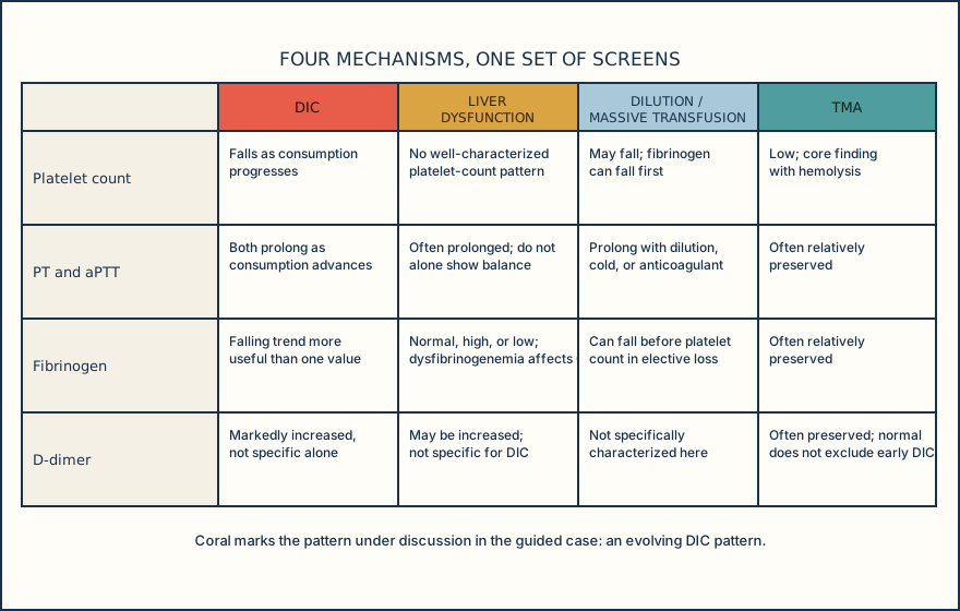
| Mechanism | Platelet count | PT / aPTT | Fibrinogen | D-dimer |
|---|---|---|---|---|
| DIC (evolving consumption) | Falls with consumption | Both prolong as consumption advances | Falling trend more informative than one value | Markedly increased, not specific alone |
| Liver synthetic dysfunction | Not directly affected by reduced factor synthesis | Often prolonged; does not alone show hemostatic balance | Normal, increased, or decreased; dysfibrinogenemia can distort Clauss result | May be increased; not specific for DIC |
| Dilution / massive transfusion | May fall with dilution | Prolong with dilution, hypothermia, or anticoagulant effect | Can fall before platelet count in elective blood loss | No specific dilutional D-dimer signature |
| Thrombotic microangiopathy | Low, with concurrent hemolysis | Often relatively preserved | Often relatively preserved | Often preserved; normal does not exclude early DIC |
Knowledge checks
Reading and checks are open. Sign in only to save.
Knowledge check 1
Knowledge check 2
Section status
Finish this section
Reading and checks are open. Sign in only to save.
The module finishes after every required section is marked done and every check in those sections is correct.