Required section · Section 3 of 6
What can prolong or hide the pattern
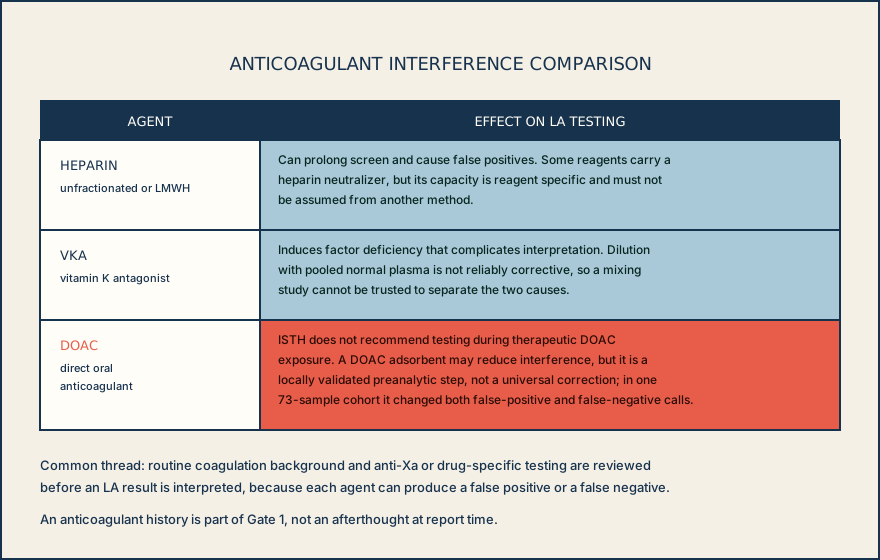
Anticoagulants are the most common confounders. Unfractionated heparin, low-molecular-weight heparin, vitamin K antagonists (VKA), and direct oral anticoagulants (DOACs) can each cause a false-positive or false-negative LA finding. Some commercial reagents contain a heparin neutralizer, but its capacity is specific to that reagent and must never be assumed to apply to a different method or lot. VKA-induced factor deficiency complicates interpretation in its own way, and dilution with pooled normal plasma is not reliably corrective for it, so a mixing study cannot be trusted to separate VKA effect from a true inhibitor.
DOACs deserve special caution because direct factor inhibition can prolong the low-phospholipid screen, persist in a 1:1 mix, and alter the high-phospholipid confirm. Those changes can resemble phospholipid dependence or hide it. Adding pooled normal plasma only dilutes drug and patient plasma together; it cannot show whether a remaining screen-to-confirm difference is caused by LA or the DOAC. Do not interpret the pattern during active therapeutic exposure. A DOAC adsorbent may reduce interference, but use it only as a locally validated preanalytic procedure for the exact assay system.
Acute-phase and specimen factors can also distort the pattern. High C-reactive protein can interfere with some phospholipid-dependent aPTT systems, while an increased factor VIII level can shorten an aPTT and obscure a weak LA rather than reveal it. On the specimen side, platelet-poor plasma matters because residual platelet phospholipid can neutralize LA and produce a false-negative result; a public accreditation checklist example specifies annual verification of residual platelet count in platelet-poor plasma with a target below 10 x 10^9/L.
Before release, confirm that the current local procedure supports the exact reagent lot, analyzer, normal-pool assignment, software algorithm, reference limits, and any neutralization claim used for the workup. When a method change has not been verified, do not carry forward the old cutoff or release an LA interpretation from it: an unverified shift in assay performance can turn anticoagulant effect or ordinary variation into an apparent inhibitor pattern.
Before you read an LA screen as positive or negative, you have already checked for anticoagulant exposure, confirmed adequate platelet-poor plasma, and noted any acute-phase flag that could change how you weigh the result.
Illustrative drawing — this picture was drawn rather than captured.
Knowledge checks
Reading and checks are open. Sign in only to save.
Knowledge check 1
Knowledge check 2
Section status
Finish this section
Reading and checks are open. Sign in only to save.
The module finishes after every required section is marked done and every check in those sections is correct.