Required section · Section 4 of 6
Working the short-fill case
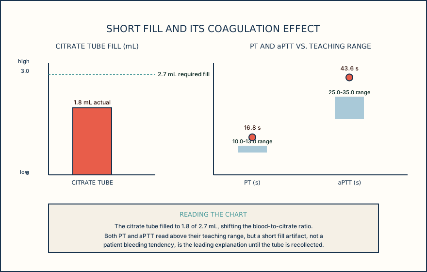
Return to the opening case. An adult outpatient has orders for blood cultures, PT, aPTT, a comprehensive metabolic panel, a complete blood count with differential, and plasma lactate. The collector uses a butterfly. The culture set is drawn first, correctly, at 09:08. The citrate tube is connected next without the local discard step, is visibly short filled at 1.8 mL against the 2.7 mL fill line, and is labeled with its actual collection time, 09:12. Serum, heparin, and EDTA tubes follow in the correct sequence. A separately collected prechilled gray lactate tube is drawn, placed on ice at 09:15, and its plasma is separated at 09:24, 12 minutes after the draw.
Work the coagulation result first. The citrate tube's short fill changes its blood-to-citrate ratio away from what the coagulation assay assumes, independent of anything happening inside the patient. Both PT at 16.8 s and aPTT at 43.6 s read above the teaching reference interval, but a short-fill artifact is the leading explanation for both, not a bleeding tendency. The hematocrit on the same accession is 43%, inside the 36 to 46% teaching interval, so the high-hematocrit adjustment pathway is not in play here; the short fill is a separate, sufficient explanation on its own.
Work the lactate specimen next. The prechilled gray tube stayed on ice from the moment of collection, and plasma was separated 12 minutes after the draw, inside the 15-minute limit from this method's example instructions. This specimen met that specific requirement, though it still needs to be checked against whatever the local test procedure for lactate specifies, because that instruction can differ by laboratory.
The defensible action on the coagulation panel is to hold release of PT and aPTT, communicate the short fill and the need for recollection through the local procedure, and retain the actual collection time and a record of that communication. The culture set's collection metadata, site, method, bottle volumes, time, and collector, must also be documented regardless of the coagulation issue, because that documentation is what makes the specimen traceable if it is ever questioned later.
A flagged coagulation result with a documented short fill is a specimen problem to resolve before release, not a patient result to interpret.
Illustrative drawing — this picture was drawn rather than captured.
| Specimen or result | Value or unit | Teaching reference or requirement | Timing or flag |
|---|---|---|---|
| PT, citrate plasma | 16.8 s | 10.0 to 13.0 s, adult interval | Short-fill flag |
| aPTT, citrate plasma | 43.6 s | 25.0 to 35.0 s, adult interval | Short-fill flag |
| Hematocrit, EDTA whole blood | 43% | 36 to 46%, adult interval | No high-hematocrit pathway |
| Lactate, plasma | Not released here | Gray tube, ice, separate within 15 min, ARUP example | Separated at 12 min |
| Blood culture set | Not released here | Collection metadata required by local SOP | Site, method, volumes, time, collector documented |
Knowledge checks
Reading and checks are open. Sign in only to save.
Knowledge check 1
Knowledge check 2
Section status
Finish this section
Reading and checks are open. Sign in only to save.
The module finishes after every required section is marked done and every check in those sections is correct.