Required section · Section 3 of 6
Use visible features and artifact checks
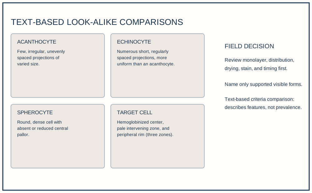
An acanthocyte has a few irregular, unevenly spaced projections. An echinocyte has many short, evenly distributed projections; each projection is distinct from a crenated edge. These comparisons are text-based criteria for review, not a recognition exercise from an image.
A qualifying schistocyte is a small RBC fragment with sharp angles and straight borders. A helmet cell qualifies when a concave missing segment forms its helmet contour; a keratocyte qualifies when two horn-like projections define the fragment. Do not count a bite cell, crenated cell, microspherocyte, or nonspecific poikilocyte as a schistocyte. A distorted cell without these criteria is excluded.
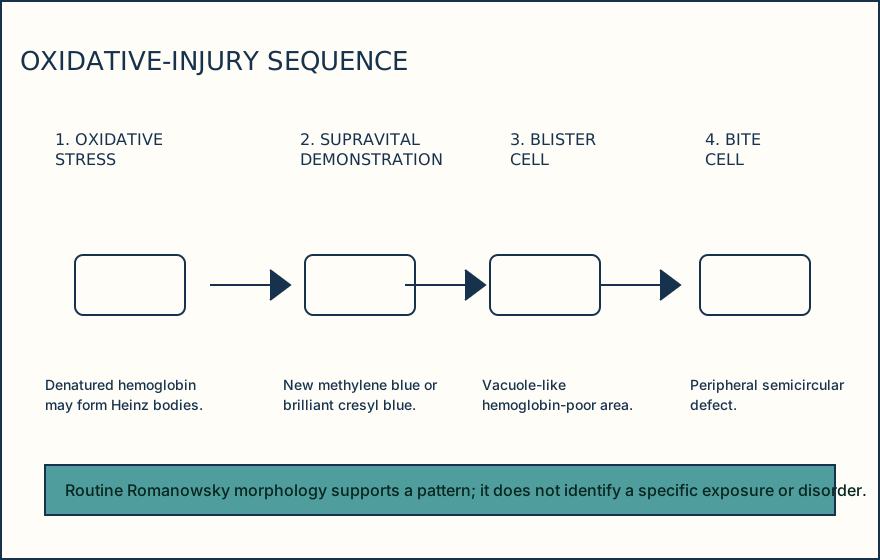
Review distribution and artifact before naming or interpreting a form. A diffuse supported bite or blister pattern with oxidative-injury context warrants the authorized oxidative review; a field-limited crenated pattern warrants preparation review or repeat smear. Romanowsky stain supports morphology; supravital staining demonstrates Heinz bodies in vitro.
Illustrative drawing — this picture was drawn rather than captured.
Illustrative drawing — this picture was drawn rather than captured.
Knowledge checks
Reading and checks are open. Sign in only to save.
Knowledge check 1
Knowledge check 2
Section status
Finish this section
Reading and checks are open. Sign in only to save.
The module finishes after every required section is marked done and every check in those sections is correct.