Required section · Section 2 of 6
Build a mechanism map
Begin with distribution and preparation, then name a form. Compare the optimal monolayer with adjacent systematic fields; a shape confined to a thick area, feathered edge, or a poorly stained region is not a released morphology comment. If distortion is diffuse or preparation is uncertain, repeat the smear or seek review before reporting the morphology.
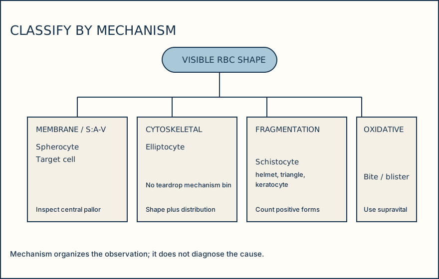
This limited comparison groups only supported visible forms. A spherocyte is round and dense with reduced central pallor. A target cell has a hemoglobinized center, a pale intervening zone, and a peripheral hemoglobinized rim. Elliptocytes are elongated. Report a supported teardrop descriptively; do not place it in a categorical mechanism bin.
Fragmentation requires positive fragment criteria. Oxidative injury can produce blister and bite forms; Heinz bodies require a separate supravital stain. Describe the observed form rather than releasing a mechanism.
Illustrative drawing — this picture was drawn rather than captured.
Evidence-first morphology workflow
Review distribution and preparation
Inspect monolayer and adjacent fields for edge concentration, drying, stain, age, or thickness artifact.
Identify supported morphology
Name only forms with defining visible features in systematic fields.
Resolve disagreement
If analyzer flag and microscopy disagree, repeat preparation or rerun/review by the local method; investigate oxidative context when bite or blister forms are supported.
Report or escalate
Release supported morphology, or hold for recollection, consultation, or unresolved-artifact suppression under procedure.
Knowledge checks
Reading and checks are open. Sign in only to save.
Knowledge check 1
Section status
Finish this section
Reading and checks are open. Sign in only to save.
The module finishes after every required section is marked done and every check in those sections is correct.