Required section · Section 4 of 6
Guided example: count the fragments
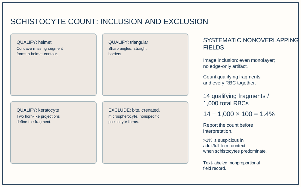
The EDTA specimen was collected at 09:10 and prepared at 09:42. After image inclusion review confirms an even Romanowsky-stained monolayer without edge-only distortion, count qualifying fragments and total RBCs together across systematic nonoverlapping fields.
In each field, record qualifying helmet, triangular, or keratocyte fragments and every RBC in the denominator before moving to the next nonoverlapping field. Across 1,000 RBCs, 14 qualifying fragments are counted: 14 ÷ 1,000 × 100 = 1.4%. Excluded bite, crenated, microspherocytic, and nonspecific distorted cells remain in the total-RBC denominator but are not numerator fragments.
Report: ‘Schistocytes 14/1,000 RBCs (1.4%) by microscopic count.’ In adults and full-term neonates, >1% morphologically identified schistocytes is suspicious for thrombotic microangiopathy when schistocytes predominate. In preterm infants, the ICSH reference upper limit is 5% schistocytes rather than the adult and full-term threshold; the adult threshold must not be transferred to a preterm result. Predominance increases specificity; it does not make a qualifying count invalid. The report is distinct from the clinical interpretation and local escalation decision.
Illustrative drawing — this picture was drawn rather than captured.
| Result | Value | Adult local interval | Method or comment |
|---|---|---|---|
| Hemoglobin | 8.6 g/dL | 12.0-16.0 g/dL | Automated CBC |
| Platelet count | 41 × 10^9/L | 150-400 × 10^9/L | Automated CBC; smear estimate consistent |
| Reticulocyte count | 6.1% | 0.5-2.5% | Optical or fluorescent method |
| LDH | 1,120 U/L | 140-280 U/L | Chemistry result for correlation |
| Schistocytes | 14/1,000 RBCs = 1.4% | >1% suspicious when predominant | Manual microscopic count using ICSH criteria |
Ordering exercise
Place the count workflow in the order that produces a standardized percentage.
1. Calculate and contextualize
Divide qualifying fragments by RBCs assessed, multiply by 100, then apply population and predominance context.
2. Identify positive forms
Count helmet, triangular, and keratocyte forms meeting criteria.
3. Select the optimal monolayer
Use a well-spread area suitable for morphology review.
4. Assess at least 1,000 RBCs
Use the denominator required for a reported percentage.
Knowledge checks
Reading and checks are open. Sign in only to save.
Knowledge check 1
Knowledge check 2
Section status
Finish this section
Reading and checks are open. Sign in only to save.
The module finishes after every required section is marked done and every check in those sections is correct.