Required section · Section 2 of 6
What each method separates
Cation-exchange HPLC separates fractions by chromatographic retention and reports relative peak areas. Its retention windows and assignments are specific to the declared analyzer, reagent, and software.
Electrophoresis separates hemoglobins by mobility. Alkaline electrophoresis can place Hb S and Hb D together; acid electrophoresis can separate that specific pair, but does not make every band uniquely identifiable.
Isoelectric focusing (IEF) separates hemoglobins by isoelectric point in a pH gradient. Capillary electrophoresis (CE) separates by electrophoretic mobility into method-specific zones. Each supplies separation evidence, not globin-gene sequence.
Molecular methods detect defined globin-gene sequence variants or copy-number changes. They can resolve an inconclusive phenotype when the selected assay covers the relevant change; sequencing alone may miss a large deletion or duplication. No one method erases the limitations of the others.
Quantitation is method output rather than a genotype statement. Reduced beta-chain production leaves a relatively greater proportion of delta-chain HbA2, so HbA2 can increase in beta-thalassemia carrier patterns. For example, co-inherited alpha-thalassemia can leave marked microcytosis with a non-diagnostic separation pattern; persistent microcytosis then follows the local molecular-referral pathway rather than being assigned from the fraction pattern.
Illustrative drawing — this picture was drawn rather than captured.
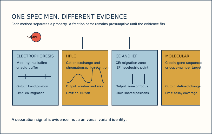
A reusable method-to-claim model for fraction review.
Name the signal
Record whether the observed evidence is a band, retention window, migration zone, isoelectric focus, or molecular target.
Name the method limit
Check whether co-migration, co-elution, assay coverage, or quantitation interference could leave more than one explanation.
Add context
Review age, CBC indices, and available transfusion, transplant, or therapy history.
Choose the next evidence
Use a complementary separation or targeted molecular method when the pattern remains unresolved or consequential.
Knowledge checks
Reading and checks are open. Sign in only to save.
Knowledge check 1
Knowledge check 2
Section status
Finish this section
Reading and checks are open. Sign in only to save.
The module finishes after every required section is marked done and every check in those sections is correct.