Required section · Section 4 of 6
Working the case: blood 2+ with a quiet sediment
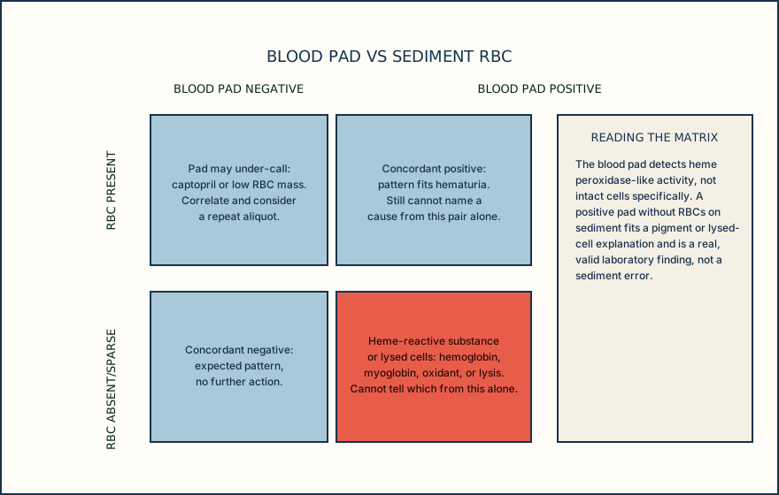
Return to the opening panel. Blood is 2+, protein is trace rather than negative, and the remaining chemistry pads and sediment findings sit within their case expectations. There is no visible red discoloration and no analyzer flag. Two sediment findings matter here: RBC is 0 to 2/HPF, within this case laboratory's local interval, and there are no casts that would suggest a glomerular or tubular process.
That combination, a positive blood pad with RBCs absent or sparse on sediment, supports a heme-reactive substance or lysed red cells more than it supports intact hematuria. It cannot distinguish hemoglobin from myoglobin, and it cannot establish either condition; distinguishing them needs additional clinical or laboratory information that this panel does not provide.
Before treating this as physiological, work the preanalytical and analytical list first. Confirm specimen identity and collection time (35 minutes is well within a reasonable window for fresh testing), confirm the strip lot has not expired and was stored correctly, confirm the day's quality control passed, and confirm the analyzer did not flag the read. Each of these is checkable from records already on file and each one is a plausible source of a blood-pad artifact.
If those checks are clean, follow the local discrepant-result algorithm with a branch matched to the evidence: repeat the strip and sediment on a fresh aliquot when a repeat can test handling or reading variation; assess RBC morphology only if the repeat sediment contains enough intact RBCs for morphology to be meaningful; use the laboratory's alternate confirmatory method when it can answer the remaining heme-reactivity question; request recollection only when specimen integrity or collection is in doubt; and communicate with the ordering clinician when a persistent, unexplained pattern needs history the laboratory does not have. None of these steps establishes a diagnosis.
Do not describe the sediment as a false negative just because the blood pad is positive; describe what each result supports and route the case through the checks the evidence, not a guess, points to.
Illustrative drawing — this picture was drawn rather than captured.
Knowledge checks
This section has no knowledge checks.
Section status
Finish this section
Reading and checks are open. Sign in only to save.
The module finishes after every required section is marked done and every check in those sections is correct.