Required section · Section 1 of 6
A blood-positive strip with a quiet sediment
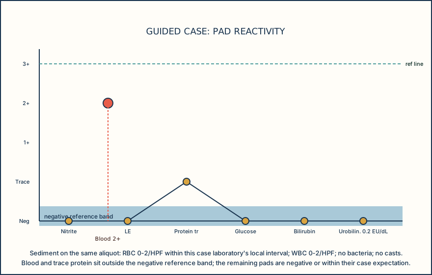
A fresh clean-catch random urine reaches the bench 35 minutes after collection, in a clean container, with no visible red discoloration. The Multistix 10 SG panel, read on a CLINITEK-family analyzer, reports blood 2+, protein trace, glucose negative, nitrite negative, leukocyte esterase negative, bilirubin negative, and urobilinogen 0.2 EU/dL. Specific gravity reads 1.015. Blood is clearly positive; protein trace is a marginal result rather than the expected negative reaction, while the remaining listed pads are within their case expectations.
The sediment from the same mixed specimen tells a quieter story: RBC 0 to 2 per high-power field (HPF), within this case laboratory's local interval; WBC 0 to 2/HPF; no bacteria; and no casts. No analyzer flag fired. If a colleague hands you this pair of reports and asks whether the blood result is wrong, the honest answer starts with what the blood pad actually measures, not with a guess.
The goal is not memorizing a rule that resolves every mismatch. It is building the habit of asking what each result can and cannot tell you, checking the variables that could produce the pattern you are looking at, and choosing a next step that a reviewer could defend from the record.
A chemistry result and a microscopy result that disagree are both still valid findings until you have a reason to distrust one of them; disagreement is a signal to investigate, not a defect to explain away.
Illustrative drawing — this picture was drawn rather than captured.
Knowledge checks
This section has no knowledge checks.
Section status
Finish this section
Reading and checks are open. Sign in only to save.
The module finishes after every required section is marked done and every check in those sections is correct.