Required section · Section 2 of 6
Chemistry and microscopy answer different questions
A urine dipstick is a screening method. Each pad is a colorimetric reaction tuned to one analyte or reaction class, read at a fixed time and compared to a color chart or an instrument's optical reader. A single pad result is a chemical measurement, not a diagnosis, and it was never meant to stand alone.
Sediment microscopy examines the formed elements in a centrifuged aliquot: intact red cells, white cells, casts, crystals, bacteria, and other structures seen and counted directly. It answers a structural question. The chemistry pads answer a chemical question. A third input, the patient's specimen collection circumstances and clinical history, frames both.
Because these three lines of evidence measure different things, agreement is common in routine specimens but is not a requirement for either result to be valid. A sediment finding does not have to confirm a chemistry finding for the chemistry to be correct, and a negative sediment does not overrule a chemically real reaction on a pad.
CAP's urinalysis checklist requires the laboratory to correlate microscopic sediment findings, such as casts, RBCs, or WBCs, with macroscopic results such as protein, occult blood, or leukocyte esterase. Correlation means the laboratory has a process for checking the two against each other, not that every specimen must show matching results before either one is released.
Before you troubleshoot a mismatch, name which of the three lines of evidence disagree and remember that each one is measuring something different from the others.
Illustrative drawing — this picture was drawn rather than captured.
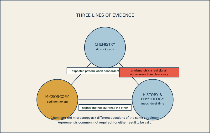
How a urine chemistry pad and a sediment field build two independent observations from the same specimen.
Collect and mix
The specimen is collected, then mixed just before testing so the aliquot used for the strip and the aliquot centrifuged for sediment start from the same suspension.
Read the chemistry pads
Each reagent pad undergoes its own colorimetric reaction at a fixed time, reflecting the chemical or enzymatic property that pad detects, and is read visually or instrumentally against expected values.
Examine the sediment
A separate aliquot is centrifuged and the formed elements, cells, casts, crystals, and organisms, are counted and described under the microscope using the local procedure.
Correlate the two reports
The laboratory compares the chemistry and microscopy results against each other and against written criteria before release, because the two methods are expected to fit a known pattern more often than not.
Knowledge checks
This section has no knowledge checks.
Section status
Finish this section
Reading and checks are open. Sign in only to save.
The module finishes after every required section is marked done and every check in those sections is correct.