
Urine Formation
Nephron Structure and Urine Formation
Every urine result reflects three nephron processes. The glomerulus filters plasma, the tubules return needed water and solutes to blood, and tubular cells add selected substances to the forming urine. Disease changes one or more of these processes. The resulting combination of protein, blood, cells, casts, crystals, concentration, and excretion provides a laboratory pattern that guides further testing.
From nephron to urine
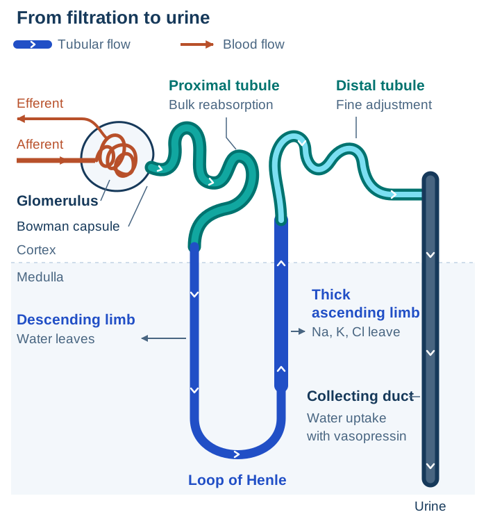
Each kidney contains about one million nephrons. Blood enters a glomerulus through an afferent arteriole and leaves through an efferent arteriole. The efferent vessel supplies peritubular capillaries around cortical tubules and the vasa recta beside long loops of Henle. The kidneys normally receive about 20% to 25% of cardiac output, with renal blood flow near 1.0 to 1.2 L/min and renal plasma flow near 600 to 700 mL/min. These are physiologic approximations that vary with body size and hemodynamic state.1,2
Filtrate enters Bowman capsule, then travels through the proximal tubule, descending and ascending limbs of the loop of Henle, distal tubule, collecting duct, renal pelvis, and ureter.
Each nephron segment has a distinct role in urine formation:
| Nephron region | Main function | Urinalysis consequence of dysfunction |
|---|---|---|
| Glomerulus | Filters water and small solutes while retaining cells and most plasma protein | Albuminuria, hematuria, dysmorphic red blood cells (RBCs), and RBC casts support glomerular barrier injury |
| Proximal tubule | Reabsorbs most filtered water and sodium, glucose, amino acids, bicarbonate, phosphate, and low-molecular-weight proteins; secretes organic acids, bases, and some drugs | Normoglycemic glycosuria, aminoaciduria, phosphate or bicarbonate wasting, low-molecular-weight proteinuria, renal tubular epithelial cells, and tubular cell casts support proximal tubular dysfunction |
| Loop of Henle | Establishes the corticomedullary osmotic gradient through countercurrent flow, NaCl transport, and urea recycling | Loss of concentrating ability produces dilute urine despite a physiologic need to conserve water |
| Distal tubule and collecting duct | Adjust sodium, potassium, water, hydrogen-ion, and ammonium excretion under hormonal control | Abnormal urine concentration or acidification can accompany vasopressin disorders, aldosterone disorders, and renal tubular acidosis |
The later renal-function lesson develops filtration control, concentrating mechanisms, function tests, and disease patterns.
References
- National Institute of Diabetes and Digestive and Kidney Diseases. Your Kidneys & How They Work. Accessed August 29, 2026.
- Strasinger SK, Di Lorenzo MS. Urinalysis and Body Fluids. 7th ed. F.A. Davis; 2021. ISBN 978-0-8036-7582-7. F.A. Davis catalog record.