The same cell count or protein result means different things in a joint, the pleural space, the pericardium, and the peritoneal cavity. Synovial, pleural, pericardial, and ascitic fluids share many laboratory methods. A high synovial leukocyte count raises concern for joint infection. Light's criteria classify pleural effusions, and the serum-ascites albumin gradient identifies the pressure mechanism behind ascites. Interpret each result for the fluid type and the clinical question.
Specimen routing and method limits
Record the anatomic site, color, clarity, volume, visible clot, and other unusual features before centrifugation or treatment. Then divide the specimen according to the tests ordered and the receiving laboratory's validated procedure. No single tube sequence or fixed aliquot volume applies to every synovial or serous fluid.
Scroll sideways to see all columns
| Test group | Specimen control |
|---|---|
| Cell count and differential | Mix promptly and use the additive, dilution, pretreatment, and analyzer mode validated for that fluid. Retain an untreated aliquot when a treatment such as hyaluronidase could affect another test. |
| Crystal examination | Examine fresh material promptly at room temperature. Use a plain or additive-containing tube only when the laboratory has shown that the container and additive do not create crystal-like material or obscure crystals. |
| Chemistry | Use the validated specimen type and method. Collect paired blood close to the fluid collection when a ratio, gradient, or direct comparison is required. |
| Microbiology | Submit a sterile aliquot with the source and requested studies. The microbiology laboratory determines volume, containers, bedside culture-bottle use, media, and special transport. |
| Cytology or flow cytometry | Protect cell morphology, submit enough specimen when possible, and follow the receiving service's instructions. Low cellularity can limit sensitivity. |
Liquid ethylenediaminetetraacetic acid (EDTA) or sodium heparin may be suitable for selected cellular methods. A plain sterile container may be preferred for others. Oxalate, lithium heparin, powdered EDTA, glove powder, and other particulate material can resemble crystals or interfere with testing. Check the laboratory's procedure for which additives it accepts. Synovial specimens require prompt analysis because cells deteriorate and crystals can precipitate or change after collection. Refrigeration before crystal examination can create in vitro crystals.1,2
Body-fluid chemistry and automated cell-count methods require verification or validation for the specific fluid matrix, analyzer mode, and intended use. Under the Clinical Laboratory Improvement Amendments, a laboratory establishes performance specifications when a method lacks applicable manufacturer claims or is modified. Those specifications include accuracy, precision, analytical sensitivity and specificity, reportable range, reference intervals, and other characteristics needed for the test system.3-5
The Body Fluid Cell Counts and Cerebrospinal Fluid lesson covers chamber selection, cytocentrifugation, analyzer limits, and morphologic review. The Manual Cell-Counting Calculations lesson covers counted volume, dilution factors, and duplicate counts.
Synovial fluid formation
Synovial fluid occupies movable joints and reduces friction between articular surfaces. Plasma ultrafiltrate crosses the synovial microvasculature, and synoviocytes add hyaluronate and other macromolecules. Type A synoviocytes have macrophage-like functions, and type B synoviocytes produce hyaluronate and matrix proteins. Hyaluronate gives healthy fluid its viscosity, and dissolved nutrients support avascular cartilage.1,2
Healthy synovial fluid is usually clear and colorless to pale yellow, has high viscosity, contains no pathologic crystals, and commonly has fewer than 200 WBCs/µL. Mononuclear cells predominate. Exact volume, protein, glucose, viscosity, cell count, and differential intervals vary with the joint, collection, method, and laboratory.1,2
Inflammation fragments hyaluronate and permits more plasma proteins and cells to enter the joint. The fluid may become cloudy, darker yellow, and less viscous. Blood can reflect trauma, anticoagulation, a vascular lesion, or hemorrhage into the joint. Uneven streaks of blood can support a traumatic aspiration. Appearance alone cannot establish the source.
The string test estimates viscosity by observing how far fluid extends before the strand breaks. Collection technique and temperature affect the result, and viscosity has limited diagnostic value. The historical Ropes mucin-clot test exposes hyaluronate to acetic acid. It is rarely useful for arthritis classification. Acetic-acid diluents also precipitate synovial mucin, so they are unsuitable for synovial cell counts.2
Cell patterns in joint disease
Scroll sideways to see all columns
| Pattern | Common appearance | WBC pattern | Interpretation limits |
|---|---|---|---|
| Normal | Clear, colorless to pale yellow, viscous | Commonly <200/µL with mononuclear predominance | Use a joint- and method-appropriate interval. |
| Noninflammatory | Clear to slightly cloudy, yellow, often retains viscosity | Commonly <2,000/µL | Osteoarthritis and other degenerative processes often fit this range. |
| Inflammatory | Cloudy yellow with reduced viscosity | Often 2,000–50,000/µL with increased neutrophils | Autoimmune and crystal disease can exceed 50,000/µL. |
| Septic | Cloudy or purulent, sometimes yellow-green | Often >50,000/µL with neutrophil predominance | The count can be lower after antibiotics, early in disease, with immune suppression, or with selected organisms. |
| Hemorrhagic | Red or blood-streaked | RBC-rich; WBC and differential can be distorted by peripheral blood | Interpret with the aspiration, peripheral counts, clinical setting, and local correction policy. |
A synovial WBC count above 50,000/µL raises the probability of septic arthritis. It does not establish infection. A count below 25,000/µL lowers the probability, and infection remains possible. No single polymorphonuclear neutrophil (PMN) percentage or lymphocyte cutoff safely separates infection from another inflammatory process. Crystal arthritis and septic arthritis can occur in the same joint.6
When specimen volume is limited and septic arthritis is suspected, the septic arthritis in native joints guideline prioritizes bacterial identification, WBC count with PMN percentage, and crystal examination. Gram stain has limited sensitivity, so a negative stain cannot exclude infection. Culture remains the main method for bacterial identification.6
Viscous fluid may require a validated hyaluronidase procedure before counting. Fibrin, fat, cartilage, tissue fragments, debris, and crystals can interfere with automated body-fluid modes. Analyzer flags, results near the lower measurement limit, unexpected counts, and atypical cells require manual confirmation or expert review according to laboratory policy.2,5
Named cellular findings such as LE cells, ragocytes, Reiter cells, rice bodies, and hemosiderin-containing macrophages have limited specificity. Report the observed morphology and clinical limitation. Suspicious cells require review by qualified personnel and may lead to cytopathology, flow cytometry, imaging, or tissue evaluation.1,2
Crystal examination
Compensated polarized-light microscopy is the main laboratory method for examining monosodium urate (MSU) and calcium pyrophosphate (CPP) crystals. Mix fresh fluid gently and examine it as a wet preparation. Ordinary light helps locate cells and crystals. Crossed polarizers show birefringence, and a first-order red compensator identifies the observed sign. Report the crystal type and whether the crystals are intracellular, extracellular, or both.2,7,8
Scroll sideways to see all columns
| Crystal or particle | Bright-field appearance | Compensated polarized-light finding | Main interpretation |
|---|---|---|---|
| MSU | Slender needles, free or within leukocytes | Strong negative birefringence: yellow when parallel to the compensator slow axis and blue when perpendicular | Supports gout |
| CPP crystal | Rhomboids or short rods, often intracellular | Weak positive birefringence: blue when parallel to the slow axis and yellow when perpendicular | Supports calcium pyrophosphate deposition disease |
| Cholesterol | Large, flat plates with notched corners | Optical descriptions differ among references; see the qualification below | Can accompany chronic effusions or inflammation |
| Corticosteroid | Variable plates or shards | Variable birefringence | Recent intra-articular injection can mimic pathologic crystals |
| Calcium oxalate | Bipyramids or envelope-like forms | Birefringent | Can occur with oxalosis or advanced kidney disease |
| Basic calcium phosphate | Very small aggregates that routine polarized microscopy usually cannot resolve | Nonbirefringent | Specialized methods such as alizarin red S or reference-laboratory analysis may be needed |
Cholesterol plates are recognized by their notched shape. Published optical descriptions conflict: one set of synovial-fluid recommendations describes nonbirefringent plates, and other morphology descriptions report birefringence. Published descriptions do not establish a consistent birefringence strength or compensator sign. Document the crystal form and optical setup, and use qualified morphology review and the laboratory's validated procedure for identification.2
The paired source image below uses conventional compensated polarized-light microscopy in the magenta column. Its gray locating column fuses nonpolarized bright-field information with single-color computational polarization and keeps the same field and crystal positions.
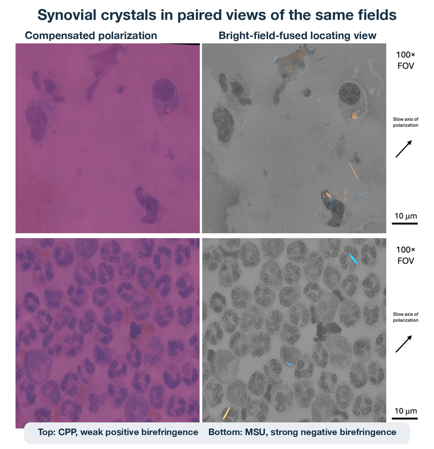
Weak CPP crystals are easy to miss, so negative microscopy does not exclude calcium pyrophosphate deposition disease. Serum urate can be normal during an acute gout attack, and hyperuricemia alone does not establish gout. Age, cartilage calcification, hemochromatosis, hyperparathyroidism, and hypomagnesemia can support a CPPD evaluation. Finding crystals does not end the infection workup when septic arthritis is clinically possible.6-8
Synovial chemistry and infection testing
Synovial chemistry provides supporting evidence. Glucose is most useful when synovial fluid and blood are collected close together. In healthy fluid, glucose is commonly within about 10 mg/dL (0.6 mmol/L) of the paired blood result. Larger decreases occur in inflammatory and infectious disease, and the ranges overlap. Prompt separation and analysis limit continued cellular glycolysis. Protein rises in many inflammatory conditions and rarely identifies the cause. Synovial lactate can increase the probability of bacterial arthritis in selected settings. Assay-dependent cutoffs and limited sensitivity make it unsuitable as a stand-alone test. Uric acid may help when crystal examination is unavailable or inconclusive.2
When infection is suspected, testing commonly includes Gram stain and aerobic and anaerobic culture. The microbiology laboratory chooses containers, culture bottles, media, incubation, and organism-directed molecular tests. Selected gonococcal, mycobacterial, fungal, or Lyme investigations depend on clinical findings, epidemiology, and locally available validated assays. Laboratory evaluation for Lyme arthritis begins with serum two-tier antibody testing. Synovial PCR is an adjunct in selected cases.6,9
The Cerebrospinal Fluid and Sterile Body Fluid Cultures lesson covers organism recovery, culture systems, special media, and molecular testing in normally sterile fluids.
Serous-fluid formation
The pleural, pericardial, and peritoneal cavities have parietal membranes lining the cavity walls and visceral membranes covering the enclosed organs. A thin fluid layer lubricates the opposed surfaces. Hydrostatic pressure, plasma oncotic pressure, membrane permeability, and lymphatic drainage determine how much fluid enters and leaves each cavity.1,10
Scroll sideways to see all columns
| Disturbance | Effect | Common examples |
|---|---|---|
| Increased hydrostatic pressure | Drives more fluid out of capillaries | Heart failure, venous obstruction, salt and water retention |
| Decreased plasma oncotic pressure | Reduces capillary fluid retention | Cirrhosis, nephrotic syndrome, severe protein loss |
| Increased membrane permeability | Allows protein, cells, and fluid to enter the cavity | Infection, inflammation, malignancy |
| Impaired lymphatic drainage | Slows removal of fluid and macromolecules | Tumor, fibrosis, inflammation, thoracic-duct injury |
An effusion can involve more than one mechanism. Appearance, cell counts, chemistry, cytology, microbiology, imaging, and the clinical setting each provide different information. Serous-fluid reference intervals are limited, and results require cavity-specific interpretation.
Pleural-fluid classification
Pleural transudates usually reflect systemic pressure or protein disturbances. Exudates usually reflect disease involving the pleura. Light's criteria classify a pleural effusion as exudative when at least one criterion is met.11
Scroll sideways to see all columns
| Light criterion | Exudative result |
|---|---|
| Pleural-fluid protein ÷ serum protein | >0.5 |
| Pleural-fluid lactate dehydrogenase (LDH) ÷ serum LDH | >0.6 |
| Pleural-fluid LDH | >two thirds of the serum LDH upper reference limit |
Collect paired blood and pleural fluid close together and use comparable methods. Light's criteria favor sensitivity and can label some diuretic-treated heart-failure effusions as exudates. In that setting, a serum-to-pleural albumin gradient above 1.2 g/dL can support a cardiac or transudative mechanism. Clinical findings remain part of the classification.11
Fluid protein alone, specific gravity, cholesterol, bilirubin ratios, total WBC count, and appearance can support an interpretation. They are not part of Light's criteria. Reserve Light's criteria for pleural fluid. Pericardial fluid and ascites are classified differently.
Pleural appearance and targeted testing
Record pleural appearance before processing. Clear or pale yellow fluid can occur in a transudate or exudate. Turbidity may reflect leukocytes, organisms, cells, debris, or lipid. Centrifugation can separate cellular turbidity from persistent lipid-rich turbidity. Blood can arise from a traumatic collection, hemorrhagic effusion, or hemothorax. A pleural-fluid hematocrit above 50% of the peripheral-blood hematocrit supports hemothorax. Dilution and collection trauma affect borderline results.11,12
Choose further tests according to the fluid's appearance and the clinical question.10,11
Scroll sideways to see all columns
| Finding or question | Main laboratory approach | Interpretation |
|---|---|---|
| Milky or persistently turbid fluid | Triglyceride, cholesterol, and chylomicron testing when needed | Triglyceride >110 mg/dL supports chylothorax. A result <50 mg/dL makes chylothorax less likely. Results from 50–110 mg/dL are indeterminate, and chylomicrons clarify the diagnosis. Pseudochylothorax commonly has cholesterol >200 mg/dL, lower triglyceride, cholesterol crystals, and no chylomicrons. Poor nutrition can lower triglyceride in chylothorax. |
| Suspected pleural infection | Appearance, Gram stain, culture, pH, glucose, LDH, cell count, and imaging correlation | Frank pus establishes empyema. Neutrophil predominance supports an acute process. It is not organism-specific. |
| Suspected tuberculosis | Adenosine deaminase (ADA), mycobacterial studies, and tissue sampling as indicated | ADA near or above 40 U/L can support tuberculosis. Local prevalence, age, immune status, assay, empyema, lymphoma, and other causes change its predictive value. |
| Suspected pancreatic or esophageal source | Pleural amylase with clinical and imaging correlation | Increased amylase is nonspecific and requires source evaluation. |
| Suspected malignancy | Cytology, cell block, immunohistochemistry, flow cytometry, or tissue biopsy as indicated | Reactive mesothelial cells can resemble malignancy. Negative cytology has limited sensitivity and cannot exclude malignancy. |
Collect pleural fluid for pH in an anaerobic blood-gas syringe without introducing air or local anesthetic contamination. Expel excess liquid heparin, cap the syringe, and analyze the sample promptly on a blood-gas analyzer validated for pleural fluid. Heparin and local anesthetic tend to lower pH. Air and delay tend to raise it.11,12
For a nonpurulent parapneumonic effusion, pH at or below 7.20 identifies high risk. Report it promptly under the laboratory's procedure. A result above 7.20 but below 7.40 is intermediate and requires assessment of LDH, glucose, imaging findings, fever, effusion volume, and septation. In this interval, LDH above 900 IU/L adds concern. A pH of 7.40 or higher indicates lower risk under this framework. If immediate pH measurement is unavailable, glucose below 3.3 mmol/L (approximately 60 mg/dL) provides less specific supporting evidence. The clinical service integrates the laboratory pattern with imaging and procedural safety and determines whether drainage is indicated.11
Pleural differentials describe the inflammatory pattern. Neutrophils commonly accompany acute infection, infarction, or pancreatitis. Lymphocyte-predominant effusions occur with tuberculosis, malignancy, and several chronic inflammatory disorders. Eosinophils at 10% or more are a descriptive finding associated with air, blood, medications, infection, and other causes. None of these patterns identifies a specific cause. Suspicious cells require cytopathology review.10,11
Pericardial-fluid interpretation
The normal pericardial cavity contains approximately 10 to 50 mL of fluid. Inflammation, infection, malignancy, trauma, uremia, autoimmune disease, systemic venous pressure, and impaired lymphatic drainage can produce an effusion. Rapid accumulation can compress the heart and contribute to cardiac tamponade.13
Pericardial protein, LDH, glucose, appearance, and cell counts have no universal intervals that reliably classify an effusion. Do not apply Light's criteria to pericardial fluid, because they can misclassify it. Testing follows the suspected cause and can include cell count and differential, bacterial culture, selected fungal or mycobacterial studies, and cytology. Neutrophil predominance supports acute inflammation or bacterial infection without proving the cause. Cytology can confirm malignant pericardial involvement. A negative result does not exclude it. Routine tumor markers and broad viral testing do not replace cause-directed evaluation.13
Ascitic-fluid interpretation
Ascites is excess fluid in the peritoneal cavity. Portal hypertension from cirrhosis is a common cause. Other causes include cardiac disease, malignancy, infection, pancreatic or biliary leakage, and lymphatic obstruction. Use the serum-ascites albumin gradient (SAAG), calculated from serum and ascitic samples collected at about the same time, to classify the pressure mechanism.14
SAAG = serum albumin − ascitic-fluid albumin
Scroll sideways to see all columns
| SAAG pattern | Main interpretation |
|---|---|
| ≥1.1 g/dL | Strongly supports portal hypertension. The cause can be hepatic, cardiac, or another portal-pressure disorder. |
| <1.1 g/dL | Suggests a process without portal hypertension, such as peritoneal malignancy, tuberculosis, pancreatic disease, or another inflammatory cause. |
A high SAAG reflects portal pressure. It does not identify a specific organ disease. Ascitic total protein further narrows the likely cause. A high SAAG with protein above 2.5 g/dL supports cardiac ascites more strongly than uncomplicated cirrhotic ascites.14
The cellular threshold for spontaneous bacterial peritonitis (SBP) is expressed as an absolute PMN count, so a percentage alone cannot be used to apply it. A count of at least 250 PMNs/µL meets the commonly used threshold in the appropriate clinical setting. For a bloody specimen, a common correction subtracts one PMN for every 250 RBCs when the laboratory's policy permits it. Culture-negative neutrocytic ascites remains clinically significant.14
SBP occurs without a surgically treatable intra-abdominal source. Secondary peritonitis arises from a perforation, abscess, inflamed organ, or another intra-abdominal source. Multiple organisms, persistently high PMNs, low glucose, high LDH, high protein, imaging, and the clinical course can raise concern for a secondary process. This laboratory pattern warrants prompt communication to the clinical service. Culture specimens should be obtained before antimicrobial treatment when possible and routed according to the microbiology procedure.14
Appearance directs follow-up testing. Cloudiness can reflect infection, malignancy, chyle, or abundant cells. Milky fluid prompts triglyceride testing and, when needed, chylomicron testing. Green or brown fluid can prompt paired bilirubin testing for bile leakage. Ascitic bilirubin above serum, or a fluid-to-serum ratio above 1, supports leakage. Ascitic amylase supports a pancreatic or gastrointestinal source when interpreted with paired serum and imaging. Fluid creatinine above serum supports urinary leakage. When tuberculosis is suspected, testing may include ADA, mycobacterial culture, and a validated nucleic-acid test.10,14
Cytology can identify metastatic or gynecologic malignancy. Its limited sensitivity makes a negative result inconclusive. Macrophages, reactive mesothelial cells, lipid-laden cells, microorganisms, mucin, and laminated calcified psammoma bodies are descriptive findings that require correlation. Tumor markers such as carcinoembryonic antigen (CEA) and cancer antigen 125 (CA 125) have limited specificity and do not replace cytology, tissue sampling, imaging, or clinical assessment.10
The PMN correction follows the general calculation principles in Manual Cell-Counting Calculations. Analytical methods for albumin, LDH, glucose, protein, lipids, bilirubin, amylase, and creatinine follow chemistry procedures. Culture, organism identification, and molecular testing follow microbiology procedures.
Practice
References
- Keohane EM, Preston MM, Mirza KM, Walenga JM, eds. Rodak's Hematology: Clinical Principles and Applications. 7th ed. Elsevier; 2025. Elsevier catalog record. Back to text
- Jokic A, Milevoj Kopcinovic L, Culej J, Kocijan I, Bozovic M. Laboratory testing of extravascular body fluids: National recommendations on behalf of the Croatian Society of Medical Biochemistry and Laboratory Medicine. Part II: Synovial fluid. Biochem Med (Zagreb). 2020;30(3):030501. doi:10.11613/BM.2020.030501 Back to text
- Clinical and Laboratory Standards Institute. Analysis of Body Fluids in Clinical Chemistry. 2nd ed. CLSI guideline C49. Clinical and Laboratory Standards Institute; 2018. Reaffirmed 2023. CLSI C49. Back to text
- Electronic Code of Federal Regulations. 42 CFR §493.1253: Standard: Establishment and verification of performance specifications. Accessed August 30, 2026. eCFR §493.1253. Back to text
- Bourner G, De la Salle B, George T, et al. ICSH guidelines for the verification and performance of automated cell counters for body fluids. Int J Lab Hematol. 2014;36(6):598-612. doi:10.1111/ijlh.12196 Back to text
- Ravn C, Neyt J, Benito N, et al. Guideline for management of septic arthritis in native joints (SANJO). J Bone Jt Infect. 2023;8:29-37. doi:10.5194/jbji-8-29-2023 Back to text
- Richette P, Doherty M, Pascual E, et al. 2018 updated European League Against Rheumatism evidence-based recommendations for the diagnosis of gout. Ann Rheum Dis. 2020;79(1):31-38. doi:10.1136/annrheumdis-2019-215315 Back to text
- Abhishek A, Tedeschi SK, Pascart T, et al. The 2023 ACR/EULAR classification criteria for calcium pyrophosphate deposition disease. Arthritis Rheumatol. 2023;75(10):1703-1713. doi:10.1002/art.42619 Back to text
- Miller JM, Binnicker MJ, Campbell S, et al. Guide to utilization of the microbiology laboratory for diagnosis of infectious diseases: 2024 update by the Infectious Diseases Society of America and the American Society for Microbiology. Clin Infect Dis. Published online March 5, 2024. doi:10.1093/cid/ciae104 Back to text
- Milevoj Kopcinovic L, Culej J, Jokic A, Bozovic M, Kocijan I. Laboratory testing of extravascular body fluids: National recommendations on behalf of the Croatian Society of Medical Biochemistry and Laboratory Medicine. Part I: Serous fluids. Biochem Med (Zagreb). 2020;30(1):010502. doi:10.11613/BM.2020.010502 Back to text
- Roberts ME, Rahman NM, Maskell NA, et al. British Thoracic Society guideline for pleural disease. Thorax. 2023;78(suppl 3):s1-s42. doi:10.1136/thorax-2022-219784 Back to text
- Asciak R, Bedawi EO, Bhatnagar R, et al. British Thoracic Society clinical statement on pleural procedures. Thorax. 2023;78(suppl 3):s43-s68. doi:10.1136/thorax-2022-219371 Back to text
- Schulz-Menger J, Collini V, Gröschel J, et al. 2025 ESC guidelines for the management of myocarditis and pericarditis. Eur Heart J. 2025;46(40):3952-4041. doi:10.1093/eurheartj/ehaf192 Back to text
- Biggins SW, Angeli P, Garcia-Tsao G, et al. Diagnosis, evaluation, and management of ascites, spontaneous bacterial peritonitis and hepatorenal syndrome: 2021 practice guidance by the American Association for the Study of Liver Diseases. Hepatology. 2021;74(2):1014-1048. doi:10.1002/hep.31884 Back to text