A positive blood pad or a cloudy specimen needs microscopy to identify the formed elements present. Urine microscopy identifies formed elements that physical examination and reagent-strip chemistry can only suggest. The result depends on a defined specimen volume, preparation method, counted volume or field area, optical system, reporting unit, and review rule. A laboratory must verify these elements together as one method because concentration and centrifugation change particle recovery.
For the anatomic basis of these findings, see Nephron Structure and Urine Formation.
From screening to microscopy
Many laboratories use a validated reflex algorithm to select specimens for microscopy. An order for microscopy, an abnormal screening result, an instrument flag, or a clinical setting named by the local protocol may trigger review. Such settings may include pregnancy, pediatric or older-adult care, diabetes, immunocompromise, and known renal disease. The criteria belong in the laboratory procedure and should reflect the population and the performance of the screening methods.1
Use the physical and chemical results to decide what to look for under the microscope. Then compare the findings for agreement, because several causes can produce the same screening result.
Scroll sideways to see all columns
| Screening finding | Sediment findings to seek | Main limitation |
|---|---|---|
| Red or brown urine | Intact red blood cells (RBCs) | Free hemoglobin, myoglobin, and nonheme pigments can discolor urine without intact RBCs |
| Turbidity | Cells, microorganisms, crystals, mucus, or contamination | A refrigerated specimen may contain abundant precipitated crystals |
| Positive blood pad | RBCs and RBC casts | Free hemoglobin and myoglobin also produce a heme reaction |
| Positive protein pad | Cells and casts | Proteinuria can occur with little visible sediment |
| Positive nitrite | Bacteria and white blood cells (WBCs) | Nitrate availability, bladder time, and organism metabolism affect the reaction |
| Positive leukocyte esterase | WBCs, WBC casts, and bacteria | Lysed WBCs can produce esterase with few intact cells remaining |
| Glucose present | Budding yeast or pseudohyphae | Glycosuria alone provides no evidence of yeast |
The Specimen Handling and Acceptability lesson covers routine collection, transport, preservation, and rejection rules. Sediment examination uses a well-mixed specimen within its validated stability period. Delay and dilute alkaline urine promote lysis of RBCs and WBCs and loss of hyaline casts. Refrigeration can precipitate urates or phosphates that obscure other particles. Bring a refrigerated specimen to the temperature required by the laboratory procedure and document any handling limitation.
Standardized sediment examination
A conventional concentrated-sediment method uses these linked steps:
- Mix the specimen and transfer the procedure's exact starting volume. The European Federation of Clinical Chemistry and Laboratory Medicine (EFLM) guideline gives 5 to 12 mL as a conventional range. A traditional method may begin with 12 mL.
- Centrifuge at the verified relative centrifugal force (RCF) and time. A common setting is approximately 400 × g for 5 minutes. The rotor radius determines the revolutions per minute: RCF = 1.118 × 10⁻⁵ × radius (cm) × RPM².
- Remove supernatant to the defined final sediment volume in one continuous motion without disturbing the packed sediment. Resuspend the sediment completely. Dividing the starting volume by the final volume gives the concentration factor. In the 12 mL example, final volumes of 0.5 mL and 1.0 mL produce 24-fold and 12-fold concentration.
- Load a fixed volume into the specified chamber or beneath the specified coverslip. A traditional slide method may use 20 µL under a 22 × 22 mm coverslip. Keep the chamber depth, coverslip area, and settling time constant.
- Scan at low power, commonly a 10× objective and about 100× total magnification, for casts and overall distribution. Examine cells, crystals, and microorganisms at high power, commonly a 40× objective and about 400× total magnification.
- Count the validated volume or number of fields and report in the unit defined by the procedure. A traditional field method averages RBCs and WBCs across at least 10 high-power fields (HPFs) and casts across at least 10 low-power fields (LPFs). Calibrated chambers and automated systems can report particles per microliter or per liter.
The starting volume, final volume, chamber, field count, and report categories form one method. Results from a different method are comparable only after verification. Centrifugation and supernatant removal can lose RBCs and WBCs and can fragment casts. Direct counting of uncentrifuged urine in a calibrated chamber can give a more accurate cell concentration, and concentration improves detection of rare casts and renal tubular epithelial cells.1
Automated particle analyzers use flow methods, digital imaging, or both. The laboratory validates the instrument's reportable categories and sets manual-review rules for flags, unusual images, rare particles, dysmorphic RBCs, nonsquamous epithelial cells, casts, crystals, and discrepancies with the physical or chemical findings.1
Optical methods
Most unstained sediment particles have little contrast. Reduced illumination improves bright-field visibility. Phase-contrast microscopy improves detection and discrimination of low-refractive-index particles and is especially useful for hyaline casts, cell detail, and dysmorphic RBCs.
Scroll sideways to see all columns
| Method | Useful application | Limitation or control |
|---|---|---|
| Bright field with reduced illumination | Routine scan and morphology | Excess light can hide hyaline casts and ghost RBCs |
| Phase contrast | Hyaline and cellular casts, dysmorphic RBCs, epithelial cells, mucus, motile trichomonads | Halo and edge effects require trained interpretation |
| Polarized light | Birefringent crystals and lipid droplets or casts | Birefringence varies by particle type and orientation |
| Supravital stain, such as Sternheimer-Malbin | Nuclear and cytoplasmic contrast | Stain performance and color require local validation |
| Toluidine blue or an acetic-acid preparation | Nuclear detail in an ambiguous round cell | Acetic acid lyses RBCs, so use a separate aliquot |
| Oil Red O or Sudan stain | Neutral lipid in cells, droplets, or casts | Polarized light supplies different information about birefringent lipid |
A concentrated Hansel- or Wright-stained preparation can demonstrate urine eosinophils. Sensitivity and specificity are poor, no threshold confirms acute interstitial nephritis, and current EFLM guidance finds routine reporting clinically unhelpful. Targeted workflows may use Gram stain, Prussian blue, dark-field microscopy, fluorescence, or interference-contrast optics for a specific organism, pigment, antigen, antibody, or structural question. These methods follow separate validated laboratory procedures.1
Cells
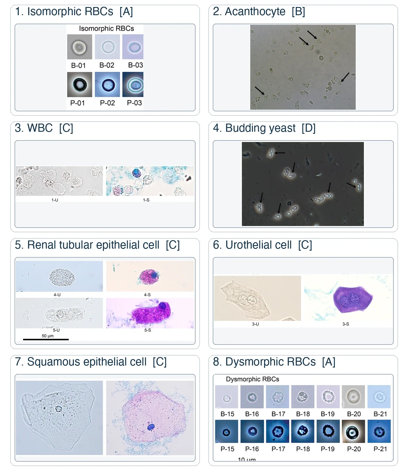
Identify a cell from its size, contour, nucleus, internal texture, and focal plane, together with specimen concentration and chemical findings. A single feature is rarely sufficient. The figure compares source photomicrographs. Preparation, stain, and optical setting vary by source and change appearance.
Red blood cells
Fresh isomorphic RBCs are smooth, nonnucleated discs approximately 6 to 7 µm in diameter. Hypersthenuric or high-osmolality urine can crenate them. Hyposthenuric or dilute urine can swell and lyse them, leaving pale ghost membranes. Fine focusing separates intensely refractile oil droplets and air bubbles from RBCs. Budding separates yeast from an RBC-like round form.1,2
Dysmorphic RBCs vary in size and shape. Acanthocytes are ring forms with multiple membrane blebs. A pattern of dysmorphic RBCs or acanthocytes in a fresh specimen supports glomerular bleeding. Phase contrast on unstained urine preserves the morphology needed for this assessment. The laboratory's validated classification requires enough RBCs for a representative percentage and correlation with protein, casts, kidney function, and the clinical setting.1,2
The 2025 American Urological Association and Society of Urodynamics, Female Pelvic Medicine and Urogenital Reconstruction guideline defines microhematuria as 3 or more RBCs per HPF in one properly collected specimen. This clinical threshold applies to a method that reports RBCs per HPF and supports risk-based clinical evaluation.3
White blood cells
Urine WBCs are generally larger than RBCs. Neutrophils are approximately 12 µm in diameter and have granular cytoplasm and a multilobed nucleus. In dilute urine, swollen neutrophils may contain granules with visible Brownian motion. These glitter cells result from hypotonic urine and have no separate clinical significance.
Pyuria can accompany bacterial infection, nonbacterial inflammation, glomerular disease, interstitial nephritis, and genital contamination. Interpret the count with collection quality, nitrite, leukocyte esterase, culture, casts, and clinical information. A WBC cast places leukocytes within a renal tubular matrix and therefore supports renal involvement.1,2
Lymphocytes, monocytes, macrophages, and renal tubular epithelial cells can appear as similar round cells. Nuclear position, cytoplasmic texture, phase contrast, and a validated stain help resolve the identity.
Epithelial cells and lipid
Scroll sideways to see all columns
| Cell | Appearance and origin | Laboratory meaning |
|---|---|---|
| Squamous epithelial cell | Very large, flat, irregular cell with abundant cytoplasm and a small central nucleus; distal urethral or genital origin | Abundant cells support collection contamination, especially with bacteria and mucus; the count alone cannot prove contamination |
| Urothelial cell | Round, pear-shaped, polygonal, or caudate cell with a central nucleus; renal pelvis, ureter, bladder, or proximal urethral origin | Small numbers can occur normally; clusters after catheterization may be reactive; atypical cells require a cytology pathway |
| Renal tubular epithelial cell | Proximal cells are large, rectangular or columnar, and coarsely granular; distal cells are smaller with an eccentric nucleus; collecting-duct cells are cuboidal with a straight edge | Increased cells or cell casts support tubular injury; cause requires clinical and laboratory correlation |
| Oval fat body | Lipid-filled renal tubular cell or macrophage with refractile droplets that may show Maltese crosses under polarized light | Supports lipiduria and heavy protein loss, especially with fatty casts and marked proteinuria |
Renal tubular cells can resemble WBCs, macrophages, or deep urothelial cells. Phase contrast or a validated stain improves nuclear detail. Prussian blue confirms hemosiderin within cells when intravascular hemolysis is under investigation. Report these advanced findings according to the laboratory's identification criteria.1,2
Clue cells in a voided urine specimen can reflect vaginal contamination. Bacterial vaginosis is evaluated with a vaginal specimen and validated diagnostic criteria.
Microorganisms, sperm, and mucus
Bacteria in a voided specimen may represent contamination, colonization, or infection. Report the microscopic finding according to the validated category and correlate it with WBCs, chemistry, collection quality, symptoms, and culture. Brownian motion can resemble bacterial motility.
Yeast appear as refractile oval forms with budding or pseudohyphae. RBCs, oil droplets, and starch can mimic yeast. Candiduria often reflects colonization, and urine findings alone cannot reliably separate colonization from infection.4
Motile Trichomonas vaginalis trophozoites are pear-shaped flagellates with an undulating membrane. Motility falls as the specimen stands. A urine wet preparation has limited sensitivity. Report a finding under the laboratory's sexually transmitted infection confirmation and reporting procedure.5
Schistosoma haematobium eggs are large, approximately 110 to 170 by 40 to 70 µm, and have a terminal spine. Travel or residence in an endemic area is essential history, and the finding requires parasite confirmation. Enterobius vermicularis eggs in urine usually reflect fecal or perineal contamination. The perianal tape test is the preferred diagnostic specimen.6,7
Spermatozoa have an oval tapered head and a long tail and are usually nonmotile in urine. They may be relevant to infertility or retrograde-ejaculation testing. Reporting depends on the specimen, patient population, and laboratory policy. Mucus forms long, pale, irregular threads and is usually insignificant. Its tapered or frayed ends and variable width help separate it from a cast.
Casts
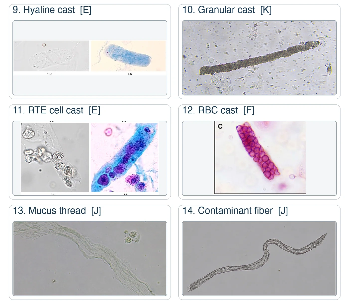
The printable cast guides show brightfield photographs and phase-contrast photographs alongside identifying features and look-alikes.
Casts form in distal tubules and collecting ducts when fibrils of uromodulin (the protein secreted by tubular cells that forms the cast matrix) gel into a cylindrical matrix. Urinary stasis, acidic pH, and favorable ionic conditions promote formation. Cells, filtered proteins, lipids, microorganisms, pigments, and crystals can become embedded in that matrix. Scan at low power to find casts, and use high power to identify their contents. A true cast has a matrix, generally with parallel sides and rounded or broken ends.1,2
Scroll sideways to see all columns
| Cast | Appearance and closest mimic | Interpretation |
|---|---|---|
| Hyaline | Colorless, homogeneous, low-refractive-index matrix; mucus has variable width and tapered or frayed ends | Can occur in concentrated urine, dehydration, or after exercise; increased counts can accompany renal disease |
| RBC | RBCs embedded within a defined matrix; free RBC clumps lack a cast outline | Strongly supports bleeding within the nephron, especially a glomerular process |
| Pigment | Homogeneous red-brown or granular matrix containing hemoglobin or myoglobin; can resemble an old RBC cast | Supports intrarenal heme pigment exposure; correlate with plasma hemolysis or muscle-injury studies |
| WBC | Leukocytes within a matrix; nuclear detail separates WBCs from renal tubular epithelial cells | Supports renal inflammation or infection and can occur with pyelonephritis, interstitial nephritis, or glomerular disease |
| Microbial | Bacteria or yeast embedded in a matrix, sometimes with WBCs; granular casts can mimic the appearance | Supports an upper-tract infectious process and requires culture or organism-specific confirmation |
| Renal tubular epithelial | Tubular cells within the matrix; degenerating cells may resemble WBCs or coarse granules | Supports tubular or renal parenchymal injury; morphology alone does not identify the cause |
| Fatty | Refractile lipid droplets or oval fat bodies in a matrix; polarization may show Maltese crosses | Supports lipiduria and heavy proteinuria |
| Granular | Fine or coarse granules within the matrix; aggregated crystals and debris can form pseudocasts | Can reflect degeneration of cells and proteins or urinary stasis; numerous muddy brown casts support tubular injury; fine versus coarse granularity has little independent clinical weight |
| Waxy | Highly refractile, brittle matrix with cracks, notches, or blunt ends | Supports prolonged stasis and advanced renal impairment |
| Broad | Wider form of another cast type | Supports formation in a dilated tubule or collecting duct; interpret the width with the cast contents and clinical findings |
| Mixed cellular | More than one cell type embedded in one matrix | Report the observed cell types and correlate with the dominant sediment pattern |
Fibers, hair, toilet tissue, mucus, and clumps of crystals can imitate a cast. Polarization, fine focus, irregular width, sharp angular edges, and the absence of a continuous matrix identify many pseudocasts. Have another qualified examiner review an ambiguous structure, following the laboratory procedure.
Crystals
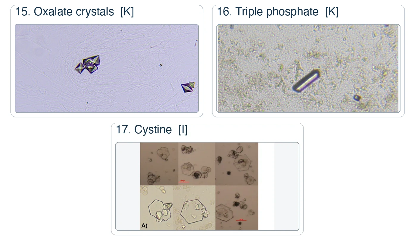
Crystals form when a substance exceeds its solubility. Urine pH, concentration, temperature, storage, diet, medication exposure, and the amount of the substance all affect precipitation. Transient crystals are common. Abundant, recurrent, atypical, drug-associated, stone-associated, or acute kidney injury-associated crystalluria requires closer identification. That identification combines shape, color, pH, polarization, solubility, medication history, and confirmatory chemistry or stone analysis.1,2
Common crystals
Scroll sideways to see all columns
| Crystal | Usual setting and morphology | Useful discriminator |
|---|---|---|
| Amorphous urates | Acidic urine; yellow-brown granules that can color refrigerated sediment pink | Gentle warming dissolves the precipitate |
| Uric acid | Acidic urine; yellow to brown rhomboids, wedges, rosettes, or plates | Strong birefringence and variable shape; hexagonal forms require distinction from cystine |
| Calcium oxalate dihydrate | Acidic to neutral urine; colorless envelope or octahedral form | Birefringent, sharply outlined envelope |
| Calcium oxalate monohydrate | Acidic to neutral urine; colorless oval, dumbbell, or elongated form | Abundant monohydrate crystals can accompany ethylene glycol exposure and require urgent clinical correlation |
| Amorphous phosphates | Neutral to alkaline urine; colorless granules that can produce a white precipitate | Dilute acetic acid dissolves the precipitate in a separate aliquot |
| Triple phosphate | Alkaline urine; colorless rectangular prisms with a coffin-lid appearance | Can accompany urease-producing organisms; chemistry and culture establish infection |
| Calcium phosphate | Neutral to alkaline urine; colorless plates, prisms, or rosettes | Acetic acid solubility and urine pH support identification |
| Ammonium biurate | Alkaline, often old urine; yellow-brown spheres with surface spicules | Strongly alkaline aged specimen supports in vitro formation |
Clinically significant and drug-associated crystals
Scroll sideways to see all columns
| Crystal or group | Morphology | Interpretation and confirmation |
|---|---|---|
| Cystine | Colorless hexagonal plates, usually in acidic urine | Supports cystinuria; confirm with amino acid analysis or another validated method |
| Cholesterol | Large flat rectangles with one or more notched corners | Correlate with oval fat bodies, fatty casts, and heavy proteinuria; see the optical qualification before assigning a polarization pattern |
| Tyrosine and leucine | Tyrosine forms fine needles in bundles; leucine forms yellow-brown spheres with concentric and radial markings | Can accompany severe liver dysfunction or an inherited metabolic disorder; confirm with current chemistry or metabolic testing |
| 2,8-Dihydroxyadenine | Round to oval brown crystals with a dark rim and radiating center | Supports adenine phosphoribosyltransferase deficiency; confirm with enzyme, molecular, or stone analysis |
| Xanthine | Variable yellow-brown granules, spheres, plates, or short rods | Can accompany hereditary xanthinuria or xanthine-oxidase inhibitor therapy; confirm with chemistry or stone analysis |
| Bilirubin | Yellow needles or granules, often in clumps | Correlate with urine bilirubin chemistry and the hepatic evaluation |
| Drug-associated crystals | Sulfonamides may form sheaves or needles; acyclovir forms long birefringent needles; indinavir can form plates or starbursts; triamterene forms yellow-brown spheres; amoxicillin can form needle bundles | Medication, dose timing, hydration, kidney function, and confirmatory analysis determine significance |
A crystal's appearance can suggest its identity. Finding one rarely establishes infection, drug toxicity, stone disease, liver disease, or an inherited disorder.
Contaminants and artifacts
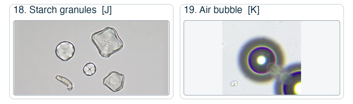
Artifacts are often highly refractile or appear in a different focal plane from the sediment. Their shape, optical behavior, and the surrounding chemical and microscopic findings guide exclusion.1,2
Scroll sideways to see all columns
| Artifact | Appearance | Distinction from a formed element |
|---|---|---|
| Starch granule | Round, refractile particle with a central dimple; Maltese-cross pattern under polarized light | Variable size and a central indentation distinguish it from RBCs; surrounding lipid findings distinguish it from oval fat bodies |
| Oil droplet or air bubble | Highly refractile circle; an air bubble often has a thick or double outline | Changes sharply with fine focus and may lie above the sediment plane |
| Pollen | Large round structure with a patterned wall, pores, or concentric detail | Size and regular wall pattern separate it from cells |
| Fiber or hair | Long, bright structure with irregular width, twists, or frayed ends | Lacks a continuous cast matrix and often polarizes strongly |
| Fecal or plant material | Coarse brown debris, plant cells, fibers, or food material | Collection history and mixed contamination support the source; unexpected fecal material requires review |
| Crystal aggregate | Angular granules or plates aligned into a cast-like clump | Individual crystal edges and polarization remain visible; no uromodulin matrix surrounds the group |
Leave artifacts out of the patient result. A mismatch between microscopy, physical appearance, and chemistry prompts review of mixing, chamber loading, focus, specimen identity, and the possibility of contamination.
Reporting and interpretation
Report the method's unit and category, the particle identity, and any permitted qualifier. Quantitative cell and cast results support comparison over time only when the specimen type and method remain comparable. Crystals, microorganisms, mucus, and artifacts often use validated ordinal categories because visual microscopy has limited counting precision for these particles. Common local terms include rare, few, moderate, and many, or 1+ through 4+. The procedure defines each category.
The American Society for Clinical Pathology (ASCP) Board of Certification (BOC) exam lists reference ranges of RBCs 0 to 3/HPF, WBCs 0 to 8/HPF, hyaline casts 0 to 2/LPF, and epithelial cells 0 to 5/HPF. These values are examination references. The laboratory establishes clinical reference intervals for its specimen and method. The ASCP BOC Examination Reference Ranges lesson contains the complete official table.8
Sediment patterns help localize a process. Dysmorphic RBCs and RBC casts support glomerular bleeding. WBC casts support renal inflammation or infection. Renal tubular epithelial cells and their casts support tubular injury. Fatty casts and oval fat bodies support lipiduria with heavy protein loss. Granular, waxy, and broad casts support increasing stasis or parenchymal injury. Combine each interpretation with specimen quality, chemistry, culture, kidney function, and the clinical setting before you report the pattern.
References
- Kouri TT, Hofmann W, Falbo R, et al. The EFLM European urinalysis guideline 2023. Clin Chem Lab Med. 2024;62(9):1653-1786. doi:10.1515/cclm-2024-0070 Back to text
- Cavanaugh C, Perazella MA. Urine sediment examination in the diagnosis and management of kidney disease: Core Curriculum 2019. Am J Kidney Dis. 2019;73(2):258-272. doi:10.1053/j.ajkd.2018.07.012 Back to text
- Barocas DA, Lotan Y, Matulewicz RS, et al. Updates to microhematuria: AUA/SUFU guideline (2025). J Urol. 2025;213(5):547-557. doi:10.1097/JU.0000000000004490 Back to text
- Pappas PG, Kauffman CA, Andes DR, et al. Clinical practice guideline for the management of candidiasis: 2016 update by the Infectious Diseases Society of America. Clin Infect Dis. 2016;62(4):e1-e50. doi:10.1093/cid/civ933 Back to text
- Centers for Disease Control and Prevention. DPDx: Trichomoniasis. Accessed August 29, 2026. Back to text
- Centers for Disease Control and Prevention. DPDx: Schistosomiasis. Reviewed June 7, 2024. Accessed August 29, 2026. Back to text
- Centers for Disease Control and Prevention. DPDx: Enterobiasis. Accessed August 29, 2026. Back to text
- American Society for Clinical Pathology Board of Certification. Medical Laboratory Scientist, MLS(ASCP) and MLS(ASCPi) Examination Content Guideline. Revised June 9, 2026. Accessed August 29, 2026. ASCP BOC examination content guideline Back to text