A watery stool can arrive with orders for electrolytes, calprotectin, and an immunochemical occult-blood test, and each test has its own specimen requirement. Each fecal test has a different purpose. Electrolytes help classify chronic watery diarrhea, leukocytes and leukocyte proteins indicate intestinal inflammation, occult-blood methods detect hemoglobin or its breakdown products, and fecal fat and pancreatic elastase assess malabsorption and pancreatic function. Collection and handling must match the method because one stool specimen may be suitable for one test and unacceptable for another.1
Stool formation and composition
Stool contains water, intestinal bacteria, undigested dietary material, sloughed epithelial cells, gastrointestinal secretions, electrolytes, and bile-pigment products. A typical adult passes about 100 to 200 g/day, although diet and transit time cause wide variation. A measured output above 200 g/day is a conventional objective threshold for diarrhea. Stool consistency and the patient's baseline pattern are also considered because clinically important loose stools can occur below that weight.1,2
Intestinal bacteria convert bilirubin derivatives to colorless urobilinogens and stercobilinogen, which oxidize to the brown pigments of normal stool. Colonic fermentation of unabsorbed carbohydrate produces gas and short-chain fatty acids. Gas can make stool float or appear foamy. Either appearance can also occur without disease.
Laboratory patterns in diarrhea
Acute diarrhea generally lasts less than 14 days. Chronic diarrhea lasts at least 4 weeks. Laboratory findings can place a specimen into a watery, fatty, or inflammatory pattern, although mixed patterns are common.1,3
Scroll sideways to see all columns
| Pattern | Findings that support it | Main limitation |
|---|---|---|
| Watery | Fecal sodium and potassium, calculated osmotic gap, stool weight, fasting response, and sometimes pH | Secretory and osmotic mechanisms can occur together |
| Fatty | Greasy or oily appearance, increased fecal fat, or a low fecal elastase result in the right specimen | Appearance alone cannot confirm steatorrhea or identify its cause |
| Inflammatory | Gross or occult blood, fecal leukocytes, calprotectin, or lactoferrin | These findings show inflammation or bleeding without identifying the cause |
Watery diarrhea reflects net intestinal secretion, retention of a poorly absorbed solute, reduced absorptive capacity, rapid transit, or a combination of these mechanisms. Osmotic diarrhea usually improves during fasting because the poorly absorbed dietary solute is removed. Secretory diarrhea often continues during fasting and may produce a large 24-hour volume. These observations guide test selection and do not establish a diagnosis.2
Stool electrolytes and the osmotic gap
Measure sodium and potassium in stool water when the clinical question is whether chronic watery diarrhea has a secretory or osmotic pattern. Calculate the gap with an assumed luminal osmolality of 290 mOsm/kg:2
Stool osmotic gap (mOsm/kg) = 290 − 2 × (stool sodium + stool potassium)
Use the assumed 290 mOsm/kg value in the calculation. Measured fecal osmolality changes after collection as bacteria continue to metabolize substrate. Its separate use is to investigate dilution, contamination, or a storage problem. A calculated gap below 50 mOsm/kg supports a secretory pattern, and a gap above 75 mOsm/kg supports an osmotic pattern. Values between those cutoffs are indeterminate. Report the gap with the interpretive limits your laboratory has validated.2
A stool specimen contains 30 mmol/L sodium and 50 mmol/L potassium:
290 − 2 × (30 + 50) = 130 mOsm/kg
The 130 mOsm/kg result is above 75 mOsm/kg and supports an osmotic contribution. Stool pH below about 5.5 can support carbohydrate fermentation. Carbohydrate malabsorption does not always produce an acidic result. Added water, urine contamination, delayed processing, and an incomplete collection can mislead interpretation, so check the specimen before you report the gap.
Steatorrhea and malabsorption
Steatorrhea is fecal fat excretion above the method-specific upper reference limit. Maldigestion limits the breakdown or solubilization of nutrients, as in exocrine pancreatic insufficiency or inadequate bile delivery. Malabsorption limits transport across the intestinal mucosa, as in celiac disease or extensive small-bowel disease. Lymphatic obstruction can also increase fecal fat.1
Pale, bulky, greasy, oily, or foul-smelling stool may suggest steatorrhea. Floating and foaming are nonspecific. Confirm increased fat with a validated qualitative or quantitative method. The historical D-xylose absorption test cannot localize mucosal disease reliably because renal function, gastric emptying, bacterial overgrowth, transit, age, and collection quality affect the result. Current evaluations usually target the suspected pancreatic, mucosal, or biliary cause directly.
Specimen collection and handling
The collection device, diet, timing, transport, and preservative come from the requested test and its current instructions.4
Scroll sideways to see all columns
| Test | Specimen requirement |
|---|---|
| High-sensitivity guaiac occult blood | Apply samples to the manufacturer's cards from the required number of bowel movements. A bulk random stool is unsuitable for screening |
| Fecal immunochemical test (FIT) | Collect with the supplied sampling probe and buffer; the number of samples and stability interval are assay-specific |
| Calprotectin, lactoferrin, or qualitative fat | Use the container, minimum quantity, temperature, and stability interval validated for that assay |
| Pancreatic elastase | Submit a semisolid or solid random stool; watery stool can dilute elastase and produce a falsely low concentration |
| Ova and parasite examination | Collect stool without urine, water, soil, or disinfectant; process or preserve it promptly with the system required by the laboratory |
| Quantitative fecal fat | Collect the entire timed specimen during the prescribed fat-controlled diet; record the collection duration |
The travel, exposure, immune-status, outbreak, and duration history determine whether broad ova and parasite testing is needed. For adults with chronic watery diarrhea and no high-risk travel or recent immigration, the American Gastroenterological Association (AGA) recommends testing for Giardia and suggests against routine examination for other ova and parasites. When microscopy is indicated, the Centers for Disease Control and Prevention advises repeat collection after an initial negative result when suspicion remains. Three specimens passed 2 to 3 days apart improve recovery.3,4
Stool is a potentially infectious specimen. Timed collections may build pressure from ongoing fermentation. Open the container carefully in the laboratory's designated work area while using the required barrier protection and splash controls.
Macroscopic examination
Record the specimen's color and consistency before sampling. Food, medication, contrast material, and transit time can mimic disease-associated appearances.1
Scroll sideways to see all columns
| Appearance | Laboratory interpretation |
|---|---|
| Brown | Usual bile-pigment color |
| Pale, clay-colored, gray, or white | Reduced bile pigment may accompany biliary obstruction; barium can produce a similar appearance |
| Black and tarry | Melena usually reflects digested blood from an upper gastrointestinal source; iron, bismuth, or charcoal can also darken stool |
| Red or maroon | Fresh lower gastrointestinal bleeding is common, and brisk upper gastrointestinal bleeding is also possible; foods, dyes, and some medications can mimic blood |
| Green | Rapid transit, diet, medication, or retained bile pigment |
| Bulky, oily, or greasy | Suggests increased fecal fat and requires laboratory confirmation |
| Watery | Diarrhea; inspect for visible blood, mucus, oil, or foreign material before aliquoting |
Visible mucus is nonspecific. Report it when the laboratory procedure requires a macroscopic description, then interpret it with the cell findings and requested microbiology studies.
Microscopic and inflammatory tests
Fecal leukocytes. Intact neutrophils support intestinal inflammation. They establish neither an infectious cause nor a specific inflammatory disease. Cells degrade during transport, occur unevenly in the specimen, and may be absent despite inflammation. The Infectious Diseases Society of America recommends against fecal-leukocyte microscopy to determine the cause of acute infectious diarrhea. Published microscopy methods and cutoffs vary too much for a universal one-to-three-neutrophil threshold.5,6
Calprotectin and lactoferrin. These leukocyte proteins remain detectable after intact cells deteriorate. AGA suggests either fecal calprotectin or fecal lactoferrin when screening adults with chronic watery diarrhea for an inflammatory condition such as inflammatory bowel disease. A calprotectin threshold near 50 µg/g is commonly used for that question. Cutoffs differ by assay and clinical setting. Infection and other intestinal inflammation can also raise either marker. Lactoferrin in human milk complicates interpretation in breastfed infants.3,5
Muscle fibers. Microscopy for striated muscle fibers is a historical screen for impaired proteolysis and rapid transit. Eosin can sharpen the striations. Count fibers with preserved cross-striations according to the laboratory's validated preparation, magnification, reading time, and reporting limit. The result has low specificity and cannot replace current evaluation of pancreatic function or intestinal disease.1
Qualitative fecal fat. Sudan III, Sudan IV, or Oil Red O stains fecal lipid orange-red. The neutral-fat preparation chiefly demonstrates triglyceride. Acidification and gentle heating convert soaps to stainable fatty acids in the split-fat preparation, which demonstrates triglyceride and fatty acids. Droplet counts depend on the stain, magnification, field size, and reading procedure. In the approximately 430× Drummey method, fewer than 100 droplets no larger than 4 µm formed the reference pattern. More than 100 droplets measuring 6 to 75 µm indicated increased fecal fat. The laboratory must report the criterion validated for its own method. Neutral-fat and split-fat patterns do not reliably distinguish pancreatic or biliary maldigestion from intestinal malabsorption.7,8
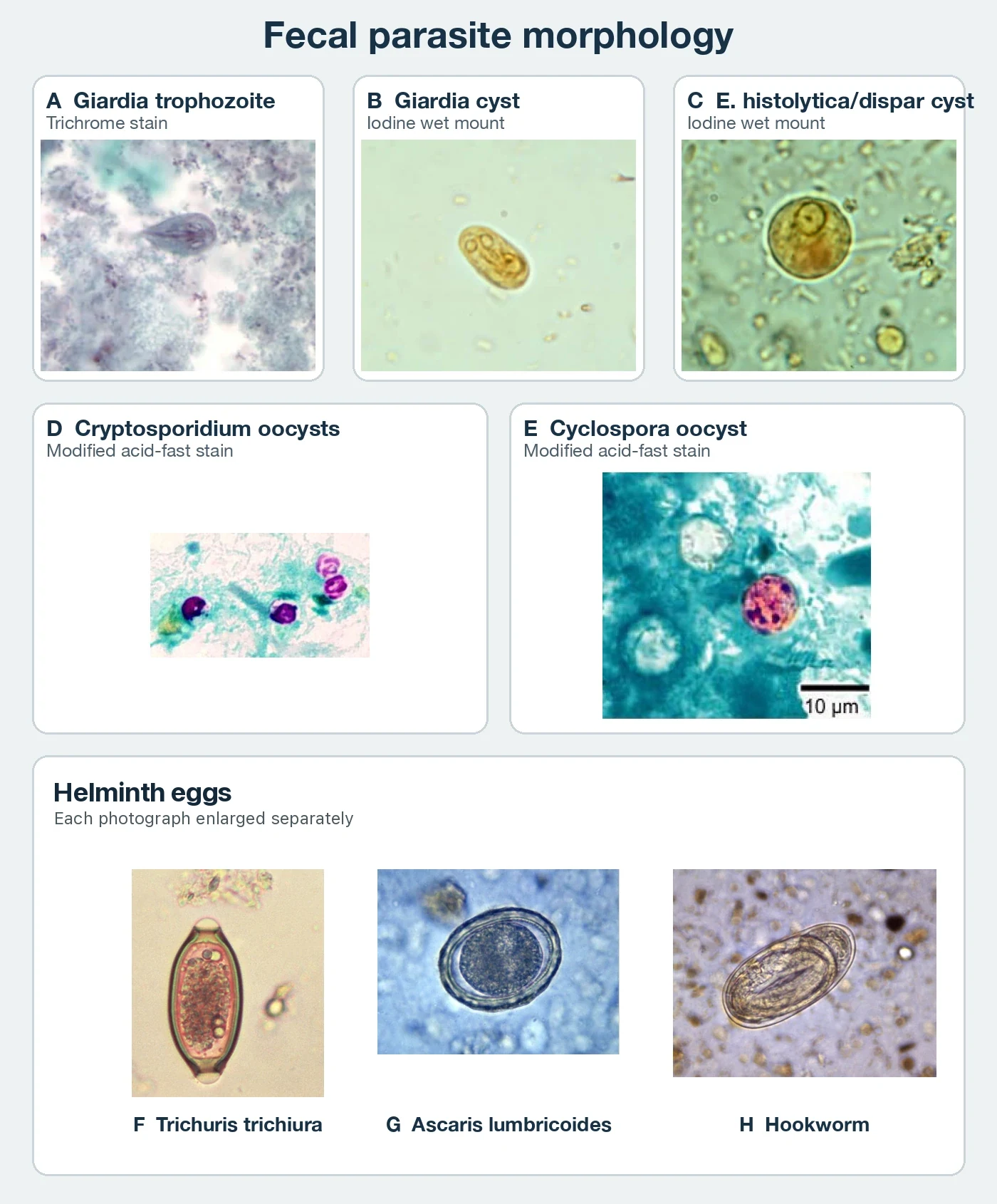
Ova and parasite examination. Promptly examined liquid stool is most likely to retain motile trophozoites. Concentration methods improve recovery of cysts, oocysts, eggs, and larvae, and a permanent stained smear supports protozoan identification. Refrigeration, preservatives, antigen tests, and molecular panels have different requirements. Store and preserve each parasite test according to the method selected for it.4
Scroll sideways to see all columns
| Parasite form | Main recognition features | Typical size |
|---|---|---|
| Giardia duodenalis trophozoite | Pear-shaped, bilaterally symmetric body with two nuclei and a ventral adhesive disc | 10 to 20 µm long |
| Giardia duodenalis cyst | Oval cyst with two nuclei when immature and four when mature | 8 to 19 µm long |
| Entamoeba histolytica/dispar cyst | Round cyst with one to four nuclei; species-level separation requires more than cyst morphology | 10 to 20 µm |
| Cryptosporidium oocyst | Round modified acid-fast oocyst, commonly staining pink to red against a blue or green background | 4 to 6 µm |
| Cyclospora cayetanensis oocyst | Round oocyst with variable modified acid-fast staining | 8 to 10 µm |
| Trichuris trichiura egg | Thick smooth shell with plugs at both poles | 50 to 55 µm long |
| Fertilized Ascaris lumbricoides egg | Thick shell, often with a mammillated outer coat | 45 to 75 µm long |
| Hookworm egg | Oval, colorless, thin-shelled egg containing a segmented embryo | 60 to 75 µm long |
Fecal occult blood methods
Fecal blood tests have different analytical targets and uses. A positive screening test identifies blood or a blood-derived product and requires diagnostic follow-up. It does not identify the bleeding site or establish cancer.10,11
Scroll sideways to see all columns
| Method | Target and main use | Important limits |
|---|---|---|
| High-sensitivity guaiac fecal occult blood test (gFOBT) | Heme pseudoperoxidase activity turns guaiac blue; used in serial colorectal-cancer screening and selected bleeding evaluations | Human and dietary heme both react; vitamin C can suppress color; collection and diet or medication instructions are product-specific |
| Fecal immunochemical test (FIT) | Antibodies detect human globin; used mainly for lower gastrointestinal bleeding and colorectal-cancer screening | Upper-tract globin may degrade before passage; cutoff, sampling device, and stability are assay-specific |
| Porphyrin quantitation (HemoQuant type) | Fluorometric measurement of heme-derived porphyrins; detects proximal and distal gastrointestinal blood loss and reports a concentration | Red meat raises results, and some porphyrias or lead intoxication can interfere; this is a specialized reference-laboratory test |
For Hemoccult II SENSA, apply two thin smears from different areas of each of three bowel movements. The product instructions prefer development at least 3 days after application so plant peroxidases can degrade on the card. If immediate testing is necessary, wait 3 to 5 minutes. Apply two drops of the matching developer over each smear and read any blue within 60 seconds. Prepared slides remain stable for up to 14 days at 15 to 30 °C. The patient instructions restrict red meat and more than 250 mg/day of vitamin C for 3 days before and during collection, and specified nonsteroidal anti-inflammatory drugs for 7 days. Medication changes require direction from the ordering clinician.10
FIT generally requires no dietary or medication restriction. For average-risk adults aged 45 to 75 years, the 2021 US Preventive Services Task Force recommendation includes annual FIT or annual high-sensitivity gFOBT among the screening options. An abnormal stool-based screening result requires colonoscopy to complete the screening process.11
HemoQuant converts intact heme and partly degraded heme products to fluorescent porphyrins. The current Mayo Clinic Laboratories method reports total hemoglobin per gram of feces and detects blood from proximal and distal sources. Its instructions call for avoiding red meat and aspirin-containing products for 3 days before collection.12
Quantitative fecal fat and acid steatocrit
Timed fecal fat confirms increased fat excretion. It does not identify the cause. The Mayo Clinic Laboratories method uses a fat-controlled diet containing 100 to 150 g/day for 3 days before and throughout collection, with 48- or 72-hour collections preferred. Laxatives, mineral or castor oil, synthetic fat substitutes, fat-blocking supplements, diaper ointments, and recent barium can interfere. Excretion above 7 g/24 h under those dietary conditions suggests malabsorption. AGA states that quantitative fecal fat is rarely needed for routine pancreatic evaluation because collection is burdensome.13,14
Report timed results as grams per 24 hours. When dietary intake has been measured, calculate the coefficient of fat absorption (CFA):
CFA (%) = [(dietary fat intake − fecal fat output) ÷ dietary fat intake] × 100
With a 100 g/day fat intake and 8 g/day fecal fat output:
[(100 − 8) ÷ 100] × 100 = 92% CFA
The calculation yields 92% CFA if the diet record and stool collection are complete. The laboratory applies the reference interval established for its population and method.13
Acid steatocrit is a rapid spot-stool screen. Acidification and centrifugation separate a fat layer from the solid layer:
Acid steatocrit (%) = fat layer length ÷ (fat layer length + solid layer length) × 100
The adult study that established a linear estimate used the following equation:
Estimated fecal fat (g/24 h) = 0.45 × acid steatocrit (%) − 0.43
That equation came from a small comparison with 72-hour fecal fat testing and is method-specific. Pediatric reference limits vary with age. A laboratory that offers acid steatocrit must validate its preparation, centrifugation, calculation, and interval.15
Apt-Downey test
The Apt-Downey test distinguishes neonatal hemoglobin from swallowed maternal blood in a fresh, visibly bloody neonatal stool or emesis specimen. Fetal hemoglobin resists alkali denaturation and remains pink after sodium hydroxide is added. Adult hemoglobin denatures to a yellow-brown product. Old, black, or tarry material may contain too little intact oxyhemoglobin for a reliable visual result.16
The current laboratory procedure sets the specimen age, reagent concentration, timing, controls, and interpretation. Increased maternal fetal hemoglobin, including hereditary persistence of fetal hemoglobin and some thalassemias, can make maternal blood appear alkali resistant and produce a misleading result.1
Pancreatic elastase
Fecal elastase is the preferred initial stool test for exocrine pancreatic insufficiency. Test a semisolid or solid specimen because watery diarrhea dilutes the enzyme and can produce a falsely low value. A result below 100 µg/g provides good evidence of exocrine pancreatic insufficiency in the appropriate setting. A result of 100 to 200 µg/g is indeterminate. The laboratory can test a specimen collected while the patient is receiving pancreatic enzyme replacement because the usual assay detects human elastase.14
Fecal chymotrypsin and trypsin methods are older indirect tests with lower clinical utility. The x-ray-film gelatin digestion screen is obsolete because intestinal degradation, trypsin inhibitors, and bacterial proteases can misclassify specimens.1
Reducing substances and carbohydrate malabsorption
Unabsorbed carbohydrate retains water in the bowel and undergoes bacterial fermentation. A copper-reduction test detects reducing sugars in stool but misses sucrose unless the specimen is hydrolyzed first. Low stool pH provides indirect evidence of fermentation. These tests performed poorly against tissue disaccharidase activity in a pediatric study, with sensitivities of 9% to 28%. Do not use them as general screening tests for disaccharidase deficiency.17
Testing should target the suspected carbohydrate and clinical setting. Options include a validated hydrogen and methane breath test or direct disaccharidase activity in an intestinal biopsy. The North American Consensus lactose breath test uses a 25 g lactose dose, measures for at least 3 hours, and defines malabsorption as a hydrogen rise of 20 ppm or more above baseline. Methane and symptoms are recorded because measured malabsorption and clinical intolerance describe different findings.18
Practice
References
- McPherson RA, Pincus MR, eds. Henry's Clinical Diagnosis and Management by Laboratory Methods. 24th ed. Elsevier; 2022. Back to text
- Schiller LR, Pardi DS, Sellin JH. Chronic diarrhea: diagnosis and management. Clin Gastroenterol Hepatol. 2017;15(2):182-193.e3. doi:10.1016/j.cgh.2016.07.028 Back to text
- Smalley W, Falck-Ytter C, Carrasco-Labra A, et al. AGA clinical practice guidelines on the laboratory evaluation of functional diarrhea and diarrhea-predominant irritable bowel syndrome in adults. Gastroenterology. 2019;157(3):851-854. doi:10.1053/j.gastro.2019.07.004 Back to text
- Centers for Disease Control and Prevention. DPDx: stool specimens, specimen collection. Accessed September 1, 2026. Back to text
- Shane AL, Mody RK, Crump JA, et al. 2017 Infectious Diseases Society of America clinical practice guidelines for the diagnosis and management of infectious diarrhea. Clin Infect Dis. 2017;65(12):e45-e80. doi:10.1093/cid/cix669 Back to text
- Babb C, Badji H, Bhuiyan MTR, et al. Evaluation of fecal inflammatory biomarkers to identify bacterial diarrhea episodes: systematic review and protocol for the Enterics for Global Health Shigella surveillance study. Open Forum Infect Dis. 2024;11(suppl 1):S65-S75. doi:10.1093/ofid/ofad652 Back to text
- Khouri MR, Huang G, Shiau YF. Sudan stain of fecal fat: new insight into an old test. Gastroenterology. 1989;96(2 Pt 1):421-427. doi:10.1016/0016-5085(89)91566-7 Back to text
- Drummey GD, Benson JA Jr, Jones CM. Microscopical examination of the stool for steatorrhea. N Engl J Med. 1961;264:85-87. doi:10.1056/NEJM196101122640207 Back to text
- Centers for Disease Control and Prevention. DPDx: stool specimens, comparative morphology tables. Accessed September 2, 2026. Back to text
- Beckman Coulter. Hemoccult SENSA and Hemoccult II SENSA Product Instructions. 462489EF. June 2015. Accessed September 1, 2026. Back to text
- US Preventive Services Task Force. Colorectal cancer: screening. Final recommendation statement. May 18, 2021. Accessed September 1, 2026. Back to text
- Mayo Clinic Laboratories. HemoQuant, feces. Test catalog. Accessed September 1, 2026. Back to text
- Mayo Clinic Laboratories. Fat, feces. Test catalog. Accessed September 1, 2026. Back to text
- Whitcomb DC, Buchner AM, Forsmark CE. AGA clinical practice update on the epidemiology, evaluation, and management of exocrine pancreatic insufficiency: expert review. Gastroenterology. 2023;165(5):1292-1301. doi:10.1053/j.gastro.2023.07.007 Back to text
- Amann ST, Josephson SA, Toskes PP. Acid steatocrit: a simple, rapid gravimetric method to determine steatorrhea. Am J Gastroenterol. 1997;92(12):2280-2284. PMID:9399770. Back to text
- Apt L, Downey WS Jr. Melena neonatorum: the swallowed blood syndrome; a simple test for the differentiation of adult and fetal hemoglobin in bloody stools. J Pediatr. 1955;47(1):6-12. doi:10.1016/S0022-3476(55)80118-7 Back to text
- Erickson JA, Case K, Neider C, Johnson LM. Stool reducing sugars and stool pH are poor screening tests for intestinal disaccharidase deficiencies in children. J Appl Lab Med. 2020;5(2):257-262. doi:10.1093/jalm/jfz017 Back to text
- Rezaie A, Buresi M, Lembo A, et al. Hydrogen and methane-based breath testing in gastrointestinal disorders: the North American Consensus. Am J Gastroenterol. 2017;112(5):775-784. doi:10.1038/ajg.2017.46 Back to text