Required section · Section 3 of 6
Focused instruction
For a person older than 12 months, cited beta-thalassemia carrier pattern data include HbA2 above 4%, HbF below 5%, and HbA above 88% (Beta-Thalassemia GeneReviews). A practical screening convention uses HbA2 greater than 3.5% (NCBI hemoglobin-disorder screening reference). Neither is a universal interval or a genotype call. Your laboratory must validate its interval and decision process on its own method.
Iron deficiency matters because severe deficiency can lower HbA2 into the normal range in a beta-thalassemia carrier. A normal or borderline HbA2 therefore does not resolve a discordant microcytic pattern. Delta-thalassemia and delta-beta-thalassemia can also alter HbA2 or HbF. Do not use one fraction to infer a beta genotype.
On this cation-exchange HPLC workflow, an HbE or Hb Lepore fraction can co-elute in the HbA2 retention window: do not report the combined peak as HbA2; inspect the chromatogram under the validated window rule and resolve it with an alternate separation method or approved reflex. In an HbS-containing specimen, HbS adducts can overlap the HbA2 window and spuriously affect integration: apply the local HbS/HbA2 integration rule and use a complementary method if the assignment remains uncertain. An alpha-chain variant can create a separate or poorly resolved abnormal peak that compromises the baseline used to integrate nearby fractions: review peak shape and baseline under the local integration rule, then use an alternate method when resolution is inadequate.
Recent red-cell transfusion can make fractionation a mixture of patient and donor cells. Newborns are different because HbF predominates at birth, and adult percentage patterns do not apply. Hb Bart greater than 15% on newborn dried-blood-spot HPLC or isoelectric focusing is suggestive of HbH disease (Alpha-Thalassemia GeneReviews), while local newborn cutoffs vary. Document age, iron studies, transfusion date, and the local validated method before interpreting HbA2 or HbF.
HbA2 is a calculated fraction: in this chromatogram assignment, the HbA2 window integrates 46,000 area units from 1,000,000 total reportable area units, so HbA2 = (46,000 ÷ 1,000,000) × 100 = 4.6%. Do not transfer this calculation to a co-eluting, overlapping, or poorly resolved window; follow the local method's assignment, review, and alternate-method pathway.
Illustrative drawing — this picture was drawn rather than captured.
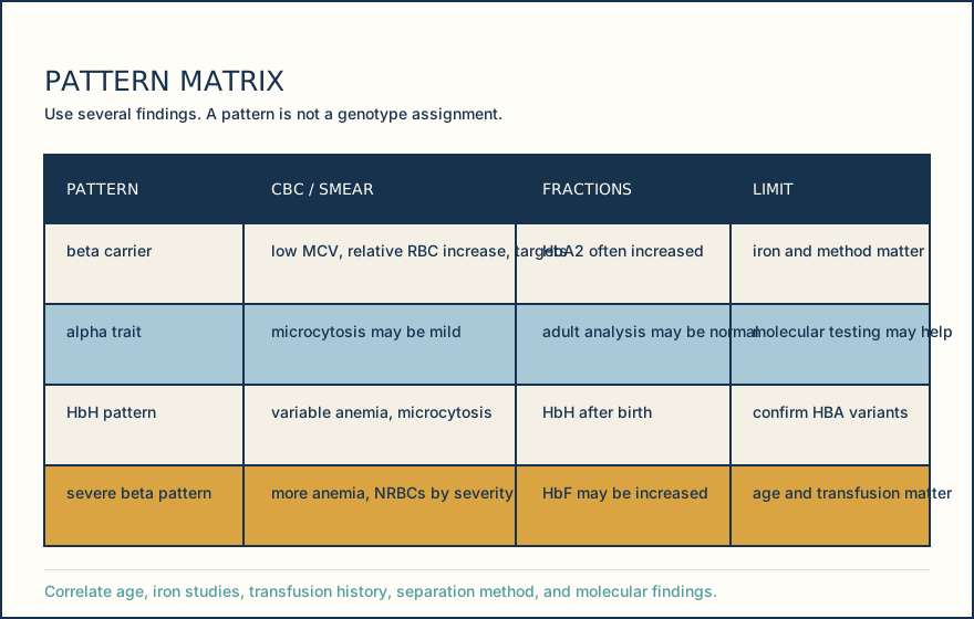
| Pattern | CBC or smear | Fraction clue | Interpretation limit |
|---|---|---|---|
| Beta carrier pattern | Microcytosis, relatively increased RBC count, target cells | HbA2 often increased | Iron status and method can alter HbA2 |
| Alpha trait pattern | Microcytosis may be present | Adult analysis may be nondiagnostic | Molecular testing may be needed |
| HbH pattern | Variable anemia and microcytosis | HbH may be detectable after birth | Confirm HBA1 and HBA2 variants molecularly |
| Severe beta pattern | More anemia, anisopoikilocytosis, nucleated RBCs by severity | HbF may be increased | Age and transfusion history alter interpretation |
Knowledge checks
Reading and checks are open. Sign in only to save.
Knowledge check 1
Knowledge check 2
Section status
Finish this section
Reading and checks are open. Sign in only to save.
The module finishes after every required section is marked done and every check in those sections is correct.