Required section · Section 3 of 6
Scanning sequence, optics, and look-alikes
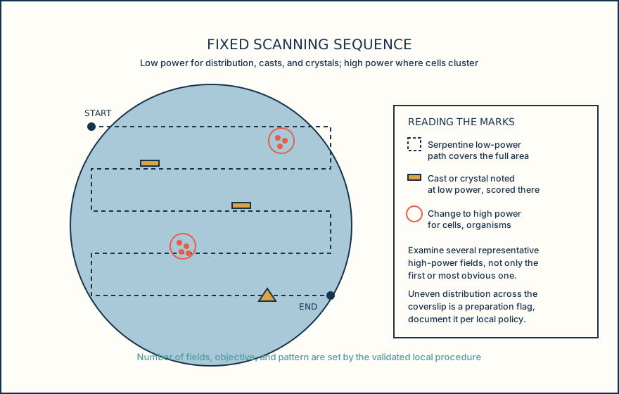
Low power is the scanning power. Its wider field is suited to surveying the whole preparation for distribution, for casts, crystals, and mucus threads that are larger or less frequent than cells, and for spotting areas where the deposit looks uneven. High power is the identification power for cells and organisms, examined in several representative fields rather than only the first one that draws the eye.
A fixed pattern, for example a serpentine path from one edge of the coverslip to the other, keeps the scan systematic instead of opportunistic. Casts and crystals flagged during the low-power pass mark where a high-power stop is worthwhile; distribution that looks patchy across the preparation is itself a finding to document, because it can mean the deposit was not mixed or loaded evenly.
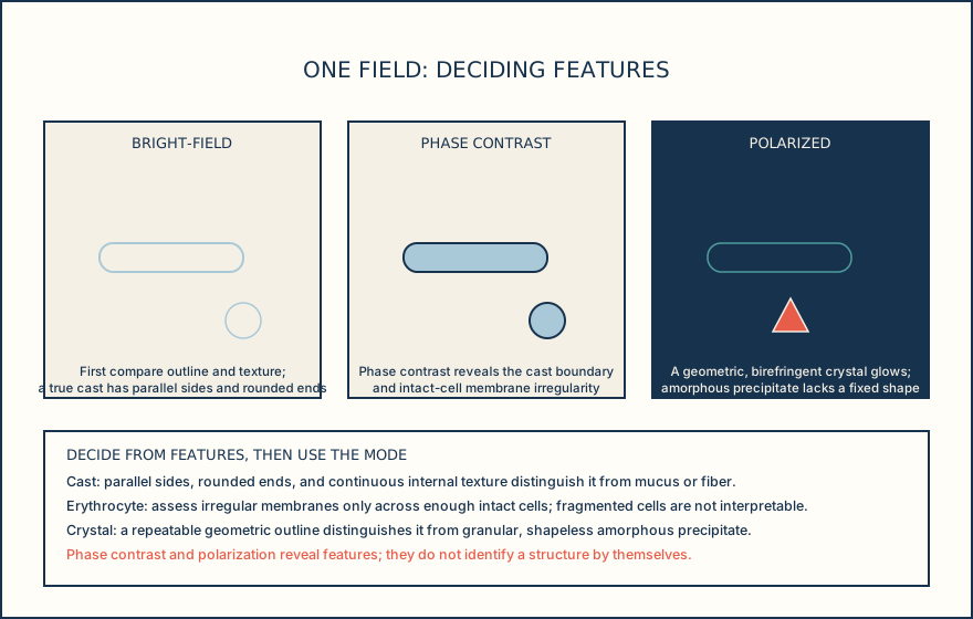
Decide an identification from visual features before choosing an optical aid. A true hyaline cast is a low-refractility structure with parallel sides, rounded ends, and a continuous internal texture; mucus threads, fibers, and scratches lack that bounded cylindrical outline or remain fixed at a different focal plane. Dysmorphic erythrocytes show membrane irregularity, blebs, or uneven contours across intact cells, whereas crenated cells have more uniform projections and fragmented cells do not support morphology assessment. A genuine crystal has a repeatable geometric outline with sharp angles or edges, unlike amorphous precipitate, which is granular and lacks a consistent shape; a starch granule can show a central hilum and a synthetic fiber has uniform width. Phase contrast can make the cast boundary and intact-cell membrane detail visible, and polarized light can test whether a candidate crystal, lipid, starch granule, or fiber is birefringent, but neither mode substitutes for the deciding features.
Field-to-volume relationships depend on the exact optical and preparation geometry: objective, field diameter, chamber depth or coverslip volume, and loaded volume. Because that geometry differs by system, a count of cells per field cannot be converted to cells per unit volume except through the laboratory's own validated system parameters, which is also why reportable units, whether per high-power field, per low-power field, a semiquantitative category, or a calculated concentration, are a local method decision rather than a universal convention.
Automated urine particle analyzers classify formed elements using a method-specific imaging or particle-recognition system and can flag a specimen for operator review. A Review status from the analyzer requires the operator to look at the flagged result; whether that step proceeds to full manual microscopy is governed by the laboratory's own validated reflex procedure, not by the analyzer flag alone. Automated categories, thresholds, and confirmation rules apply only with the exact analyzer, software version, and instructions for use the laboratory has validated.
Match the imaging mode to the specific visual ambiguity in front of you, not to a fixed task category, and treat an analyzer review flag as a prompt to look, not a verdict.
Illustrative drawing — this picture was drawn rather than captured.
Illustrative drawing — this picture was drawn rather than captured.
| Magnification | Primary use | Typical elements |
|---|---|---|
| Low power | Scan the whole preparation for distribution and larger, less frequent elements | Casts, crystals, mucus threads, distribution pattern |
| High power | Evaluate cellular detail across several representative fields | Erythrocytes, leukocytes, epithelial cells, organisms |
| Mode | What it reveals | Deciding feature |
|---|---|---|
| Bright-field | General visibility of an unstained sediment | A true cast has parallel sides, rounded ends, and continuous texture; a mucus thread or fiber does not. |
| Phase contrast | Contrast for low-refractility structures and intact-cell membranes | Confirm a cast boundary or assess membrane irregularity only across enough intact RBCs; it does not make fragmented cells interpretable. |
| Polarization | Birefringence detection | A candidate crystal has a repeatable geometric outline; amorphous precipitate is granular and shapeless. Birefringence supports, but does not replace, that comparison. |
Knowledge checks
Reading and checks are open. Sign in only to save.
Knowledge check 1
Knowledge check 2
Knowledge check 3
Section status
Finish this section
Reading and checks are open. Sign in only to save.
The module finishes after every required section is marked done and every check in those sections is correct.