Required section · Section 3 of 6
Look-alikes, motion, and the edge of routine identification
Four look-alike problems account for most of the disputed calls in routine sediment work. A partially lysed RBC can look like a yeast cell because both can appear as a pale, round-to-oval structure of similar size; the RBC has no wall and no bud, while yeast has a smooth defined wall and, when present, a narrow-based bud or pseudohyphae that are stronger evidence than color alone. A WBC and an RTE cell can overlap in size and general roundness; the RTE cell typically shows one round, centrally placed nucleus and coarsely granular cytoplasm, rather than the multilobed or degenerating nucleus of a WBC, and any RTE call still routes through the local escalation policy rather than resting on morphology alone.
Bacteria present a motion problem more than a size problem. True bacteria appear as very small cocci or rods, near the lower limit of resolution at 400x, and their apparent movement can be ordinary Brownian motion rather than organism-directed travel. Bacteriuria on a sediment must be read together with specimen collection quality, squamous epithelial cell counts, WBCs, and chemical findings, because microscopy alone does not establish a urinary tract infection; a heavily contaminated collection with many squamous cells and scattered bacteria reads differently than a clean-catch specimen with few squamous cells and the same bacterial count.
A motile, pear-shaped organism raises the question of Trichomonas-like motility. A wet-mount urine preparation is not sensitive enough to exclude Trichomonas vaginalis when the finding is negative, so a negative wet mount is evidence of absence only in a limited sense and does not clear the patient of infection. The bench response to a moving pear-shaped structure is to document the motility, examine it across multiple focus planes, compare its displacement against nearby debris to rule out simple drift, and route it through the local testing or referral pathway rather than reporting a definitive identification from the wet mount alone.
Non-biological artifacts round out the list a reader must clear before naming anything a cell or organism: air bubbles, oil droplets, starch granules, pollen, mucus threads, fibers, and occasional paper, glass, or textile debris are all established urine-sediment artifacts. Oil droplets are refractile and can resemble RBCs or yeast at first glance but lack any wall, nucleus, or bud and typically vary widely in size within the same field, which true cells rarely do. Polarized light can help characterize birefringent material when the local procedure specifies it, but it is a method adjunct, not a universal confirmation that something is a cell, bacterium, or yeast, and bright-field, phase-contrast, and polarized configurations each have a defined optical role that the local procedure must specify.
Illustrative drawing — this picture was drawn rather than captured.
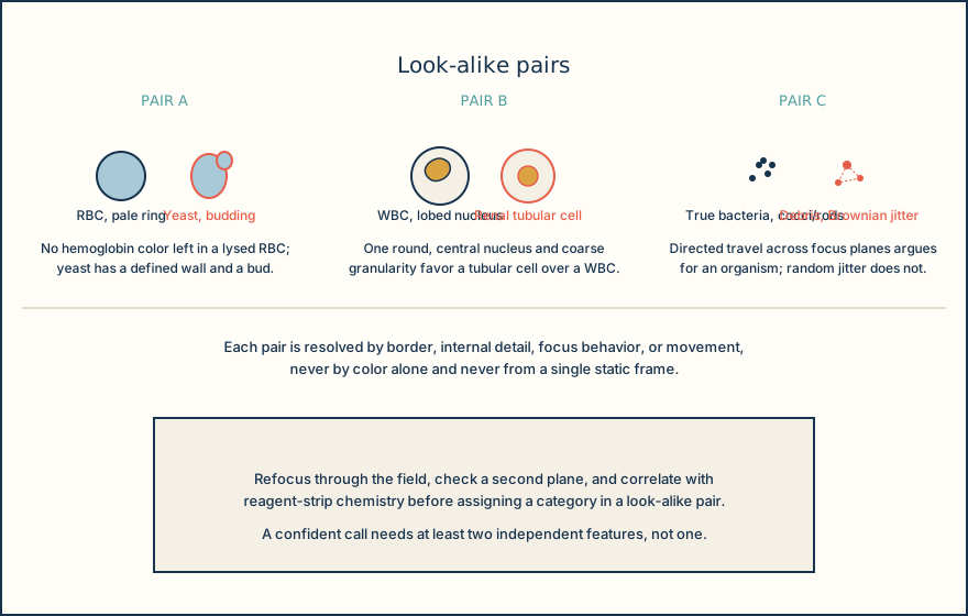
Illustrative drawing — this picture was drawn rather than captured.
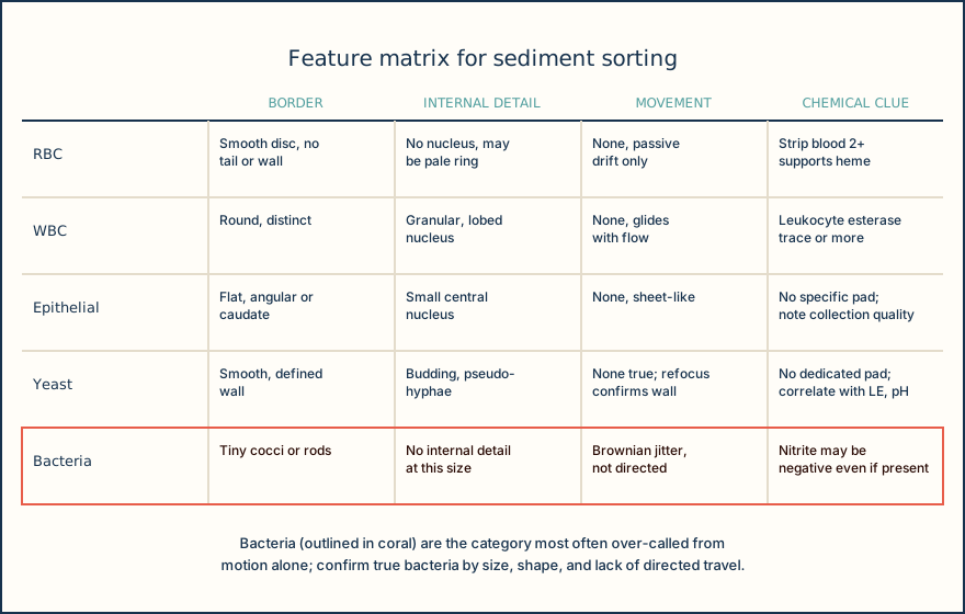
| Pair | Common trap | Resolving feature |
|---|---|---|
| Lysed RBC vs. yeast | Both round, pale, similar size to RBC | Yeast has a defined wall and may show budding or pseudohyphae |
| WBC vs. renal tubular epithelial cell | Both round with a visible nucleus, overlapping size | RTE cell has one round central nucleus and coarsely granular cytoplasm; escalate per local policy |
| True bacteria vs. Brownian debris | Both tiny and appear to move | True organism motion is directed across focus planes; Brownian motion is random jitter |
| Oil droplet vs. RBC or yeast | Refractile and rounded | Oil droplets lack a wall or nucleus and vary widely in size within one field |
Knowledge checks
Reading and checks are open. Sign in only to save.
Knowledge check 1
Knowledge check 2
Section status
Finish this section
Reading and checks are open. Sign in only to save.
The module finishes after every required section is marked done and every check in those sections is correct.