Required section · Section 2 of 6
The red cell as an internal ruler
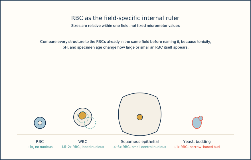
The fastest way to sort a crowded field is to use intact RBCs already present as a built-in size reference, when intact RBCs are visible in the field, because RBCs are the smallest common cellular element and appear in nearly every sediment. A structure roughly the same size as a nearby intact RBC and lacking a nucleus is a reasonable RBC candidate. A structure one and a half to two times the size of a nearby intact RBC, with a granular cytoplasm and a lobed or degenerating nucleus, behaves like a WBC. A much larger, flat structure with abundant cytoplasm and a single small central nucleus behaves like a squamous epithelial cell.
This internal-ruler approach exists because no single micrometer value can be assigned to every urine RBC. Tonicity, pH, and specimen age all change apparent RBC size and shape: RBCs can appear crenated in concentrated urine, swollen or lysed in dilute or alkaline urine, and reduced to faint membrane outlines called ghost cells after lysis. A fixed number memorized from a textbook will mislead the reader on a specimen that does not match the textbook's conditions, while a same-field comparison against intact RBCs adjusts automatically when intact RBCs are present in the field to compare against.
Urothelial (transitional) cells complicate a pure size rule because they can be round, pear-shaped, or caudate and originate anywhere from the renal pelvis through the ureters, bladder, and proximal urethra, so their shape varies more than a squamous cell's does. Renal tubular epithelial (RTE) cells add a further caution: they overlap in size and appearance with both urothelial cells and WBCs, so a confident RTE call needs cautious morphologic review and follows the validated local escalation policy rather than a size estimate alone.
None of this replaces reagent-strip chemistry results. A positive blood reaction or a positive LE result adds correlating evidence, not a cell count, and the two data streams are read together rather than forced to agree.
Illustrative drawing — this picture was drawn rather than captured.
Size-first triage workflow for an unfamiliar sediment structure.
Scan at low power
Survey the field at low power to locate clusters of cells, organisms, or debris and to confirm a well-mixed, evenly distributed preparation.
Compare size to nearby RBCs
Switch to high power and compare the unknown structure's size to intact RBCs already visible in the same field rather than to a memorized value.
Assess border and internal detail
Look for a nucleus, granularity, a defined wall, budding, or a smooth featureless border before naming the structure.
Check focus planes and movement
Rack through focus planes to separate a true organism's directed travel or a wall's contour from Brownian jitter or a flat artifact.
Correlate with chemistry
Read the reagent-strip blood and leukocyte esterase results alongside the microscopic finding, treating agreement or disagreement as information rather than a tiebreaker.
Knowledge checks
Reading and checks are open. Sign in only to save.
Knowledge check 1
Knowledge check 2
Section status
Finish this section
Reading and checks are open. Sign in only to save.
The module finishes after every required section is marked done and every check in those sections is correct.